. 2007 Feb;7(2):170-8.
doi: 10.1039/b612966h. Epub 2006 Nov 24.

A microfluidic device for practical label-free CD4(+) T cell counting of HIV-infected subjects

Affiliations

A microfluidic device for practical label-free CD4(+) T cell counting of HIV-infected subjects

Xuanhong Cheng et al. Lab Chip. 2007 Feb.

Abstract

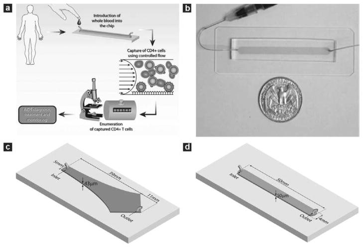
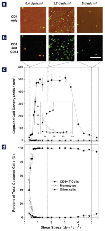
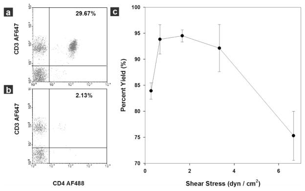
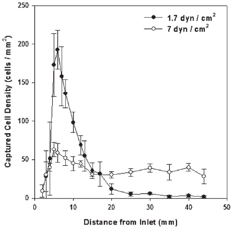
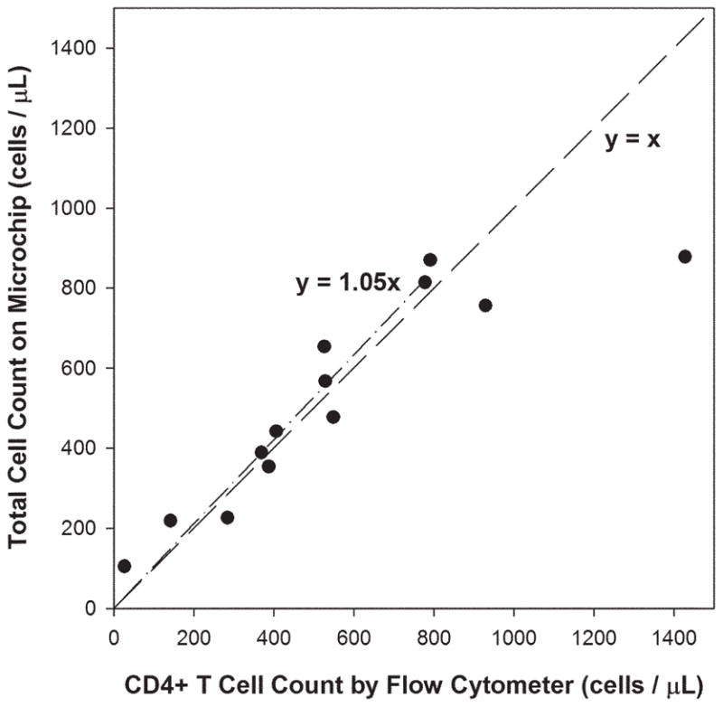
Practical HIV diagnostics are urgently needed in resource-limited settings. While HIV infection can be diagnosed using simple, rapid, lateral flow immunoassays, HIV disease staging and treatment monitoring require accurate counting of a particular white blood cell subset, the CD4(+) T lymphocyte. To address the limitations of current expensive, technically demanding and/or time-consuming approaches, we have developed a simple CD4 counting microfluidic device. This device uses cell affinity chromatography operated under differential shear flow to specifically isolate CD4(+) T lymphocytes with high efficiency directly from 10 microliters of unprocessed, unlabeled whole blood. CD4 counts are obtained under an optical microscope in a rapid, simple and label-free fashion. CD4 counts determined in our device matched measurements by conventional flow cytometry among HIV-positive subjects over a wide range of absolute CD4 counts (R(2) = 0.93). This CD4 counting microdevice can be used for simple, rapid and affordable CD4 counting in point-of-care and resource-limited settings.


Figures

Fig. 1
Fig. 1
Microfluidic devices used in the study and operating procedure of the counting device. (a) Schematic depiction showing the operating procedure of the CD4 counting device. The microchip is operated by injecting 10 μL whole blood at controlled flow rate. This is followed with rinsing unbound cells from the chamber and counting all the captured cells within the chip using an optical microscope to obtain CD4 counts. (b) Photograph of a linear cell count device. Microfabricated PDMS devices with one inlet and one outlet were bound to glass slides to form closed chambers. The chamber was functionalized with specific antibody to capture target cells from whole blood. The shaded area indicates the chamber location within PDMS. (c) Geometry of the Hele–Shaw device. The Hele–Shaw device offers a linear variation of shear along its central line. It was used in this study to screen the optimal shear stress for cell capture. (d) Geometry of the linear cell count device. The linear device has a volume of 10 μL for sample volume metering. It was operated under the optimized shear stress to capture and numerate the target cells.
Fig. 2
Fig. 2
Effect of shear stress on cell adhesion in the Hele–Shaw devices using whole blood from healthy subjects. (a) Representative images of cells captured in the Hele–Shaw chamber at locations corresponding to shear stresses of 0.4 (left), 1.7 (middle) and 5 dyn cm−2 (right). The image was created by overlapping a phase contrast photograph and the corresponding fluorescence photograph. All the cells in the phase contrast image are stained positively (green) for the CD4 surface marker, but captured cell density is greatly dependent on the shear stress. (b) Representative images of captured cells after CD4 (green) and CD14 (red) staining at the shear stresses conditions as described in (a). Both lymphocytes (CD4+CD14−, green) and monocytes (CD4+CD14+, yellow) were captured at the shear stress of 0.5 dyn cm−2, but pure lymphocytes were captured at two higher shear stresses. (Bar: 100 μm) (c) Adhesion of CD4+ T cells (solid circles), monocytes (empty circles) and other cells (solid triangles) in response to shear stress. Differentiated capture of monocytes and lymphocytes in response to shear was observed: a shear stress window between 1 and 3 dyn cm−2 was optimal for CD4+ T cell adhesion, while monocyte adhesion drops significantly above 0.7 dyn cm−2 (inset). The adhesion of other cells is minimal in the whole range of tested shear stress. (d) Composition of the surface captured cells as a function of shear stress. When shear stress is above 0.7 dyn cm−2, >95% of the surface captured cells are CD4+ T cells (solid circles). The purity of these cells drops quickly to less than 50% when shear stress drops below 0.7 dyn cm−2. In (c) and (d), each data point was repeated in 3 devices spanning different shear stress ranges; error bars represent standard deviations in measurements within each experiment.
Fig. 3
Fig. 3
Dependence of capture yield on shear stress in a linear device evaluated by flow cytometry using 10 μL blood samples from healthy subjects. (a) Flow cytometric analysis of a blood sample before CD4+ T cell isolation. The CD4+ T cells (CD3+CD4+) compose 29.67% of all lymphocytes. (b) Flow cytometric analysis of the same blood sample after CD4+ T cell capture in the device. The composition of the target cells in the sample flow through dropped to 2.13% of all lymphocyte population after device capture. Ten microlitres of whole blood were injected into the linear device at a shear stress of 1.7 dyn cm−2. Cells were acquired in the gated lymphocyte population, and the quadrants were set up with an isotype-matched control. (c) Capture yield at different shear stress calculated from flow cytometric analysis. Nearly 95% of the target cells can be isolated from whole blood using shear stress in the range of 1 to 3 dyn cm−2. The yield quickly drops to less than 85% out of this range. Each data point was repeated in at least 3 devices. The error bars represent standard deviations in measurements within each experiment.
Fig. 4
Fig. 4
Distribution of CD4+ T cells along the linear cell capture chamber at two shear stresses. At 1.7 dyn cm−2 (solid circle, yield nearly 95%), captured cell density reaches maximum near the sample inlet. By contrast, at 7 dyn cm−2 (empty circle, yield ~ 75%), the distribution of cells is fairly uniform along the device. The experiments were performed using 10 μL of whole blood from healthy subjects. Each data point was repeated in at least 3 devices. The error bars represent standard deviations in measurements within each experiment.
Fig. 5
Fig. 5
Correlation of total cell counts in the linear microchip versus absolute CD4 counts by flow cytometers, using whole blood from 13 HIV+ adult subjects. A linear regression of the experimental data for absolute CD4 counts under 800 cells μL−1 (n = 11) indicates good correlation between the two measurements (dash–dot line). The dash line represents an ideal 1 : 1 correlation between the two.
Fig. 6
Fig. 6
Purity and yield of captured CD4+ T lymphocytes in the linear cell count device using whole blood from 13 HIV-positive adult subjects. (a) Purity of the surface captured CD4+ T cells as a function of the absolute CD4 counts. Purity was above 60% and fairly consistent for absolute CD4 counts greater than 200 cells μL−1. (b) Yield of CD4+ T cells within the linear device as a function of the absolute CD4 counts. Fairly consistent yield was observed for absolute CD4 counts up to 800 cells μL−1. The dashed lines are drawn as a visual guide.

Similar articles

Cited by

References

    1. Joint United Nations Programme on HIV/AIDS. World Health Organization; Dec, 2005. [accessed 20th March 2006]. AIDS Epidemic Update: December 2005. available at http://libdoc.who.int/unaids/2005/929173439X_eng.pdf.
    1. Guidelines for the Use of Antiretroviral Agents in HIV-1-Infected Adults and Adolescents. Department of Health and Human Services; Oct 6, 2005. [accessed 20th March 2006]. available at http://aidsinfo.nih.gov/ContentFiles/AdultandAdolescentGL.pdf.
    1. Obrien WA, Hartigan PM, Daar ES, Simberkoff MS, Hamilton JD. Ann Intern Med. 1997;126:939–945. - PubMed
    1. Mellors JW, Munoz A, Giorgi JV, Margolick JB, Tassoni CJ, Gupta P, Kingsley LA, Todd JA, Saah AJ, Detels R, Phair JP, Rinaldo CR. Ann Intern Med. 1997;126:946–954. - PubMed
    1. Obrien WA, Hartigan PM, Martin D, Esinhart J, Hill A, Benoit S, Rubin M, Lahart C, Wray N, Finegold SM, George WL, Dickinson GM, Klimas N, Diamond G, ZollaPazner SB, Jensen PC, Hawkes C, Oster C, Gordin F, Labriola AM, Spivey P, Matthews T, Weinhold K, Drusano G, Egorin MJ. N Engl J Med. 1996;334:426–431. - PubMed

Publication types

MeSH terms