. 2007 Feb 1:8:39.
doi: 10.1186/1471-2164-8-39.

A screen for nuclear transcripts identifies two linked noncoding RNAs associated with SC35 splicing domains

Affiliations

A screen for nuclear transcripts identifies two linked noncoding RNAs associated with SC35 splicing domains

John N Hutchinson et al. BMC Genomics. .

Abstract

Background: Noncoding RNA species play a diverse set of roles in the eukaryotic cell. While much recent attention has focused on smaller RNA species, larger noncoding transcripts are also thought to be highly abundant in mammalian cells. To search for large noncoding RNAs that might control gene expression or mRNA metabolism, we used Affymetrix expression arrays to identify polyadenylated RNA transcripts displaying nuclear enrichment.

Results: This screen identified no more than three transcripts; XIST, and two unique noncoding nuclear enriched abundant transcripts (NEAT) RNAs strikingly located less than 70 kb apart on human chromosome 11: NEAT1, a noncoding RNA from the locus encoding for TncRNA, and NEAT2 (also known as MALAT-1). While the two NEAT transcripts share no significant homology with each other, each is conserved within the mammalian lineage, suggesting significant function for these noncoding RNAs. NEAT2 is extraordinarily well conserved for a noncoding RNA, more so than even XIST. Bioinformatic analyses of publicly available mouse transcriptome data support our findings from human cells as they confirm that the murine homologs of these noncoding RNAs are also nuclear enriched. RNA FISH analyses suggest that these noncoding RNAs function in mRNA metabolism as they demonstrate an intimate association of these RNA species with SC35 nuclear speckles in both human and mouse cells. These studies show that one of these transcripts, NEAT1 localizes to the periphery of such domains, whereas the neighboring transcript, NEAT2, is part of the long-sought polyadenylated component of nuclear speckles.

Conclusion: Our genome-wide screens in two mammalian species reveal no more than three abundant large non-coding polyadenylated RNAs in the nucleus; the canonical large noncoding RNA XIST and NEAT1 and NEAT2. The function of these noncoding RNAs in mRNA metabolism is suggested by their high levels of conservation and their intimate association with SC35 splicing domains in multiple mammalian species.


Figures
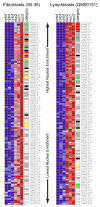
Figure 1
Figure 1
Nuclear Enriched Probe Sets in Lymphoblasts and Fibroblasts. Representative microarray hybridization of the 113 probe sets expressed above threshold that were more than 2 fold enriched in nuclear fraction samples (nuc1, nuc2 and nuc3) versus cytoplasmic fraction samples (cyto1, cyto2 and cyto3) on average in both fibroblasts (WI-38) and EBV-transformed lymphoblasts (GM00131). Probe sets are ranked in descending order of nuclear fraction enrichment from top to bottom. Probe expression is row normalized to show high expression as red and low expression as blue. Nuclear enriched probes were qualified into separate transcript type categories using the University of California at Santa Cruz genome browser and represented graphically beside the representative microarray hybridization. Probes to introns are represented in grey, protein coding transcripts (ORF containing) in white, the XIST locus in red, the NEAT1 locus in green and the NEAT2 locus in yellow.
Figure 2
Figure 2
Genomic Organization of the Human and Mouse NEAT1 and NEAT2 Loci. (A, B) Graphical representation of locations and lengths of transcripts and probes mapping to the genomic region of (A) human NEAT1/NEAT2 on 11q13.1 and (B) mouse Neat1/Neat2 on 19qA. All transcripts run 5' to 3' from left to right. Transcripts represented in green (NEAT1, NEAT2, Neat1 and Neat2) were characterized by Northern analysis, 5'RLM-RACE and 3' RACE. Genbank accession number for these transcripts are: NEAT1 – EF177379, NEAT2 – EF177381, Neat1 – EF177378 and Neat2 – EF177380. For these transcripts, all 3' poly(A) ends had a consensus AAUAAA 10–30 nucleotides 5' of the poly(A) site and a GU rich and/or U rich element 3' of the site. Human NEAT1 is observed to extend past its primary poly(A) site, resulting in a larger, >17 kb transcript. The 3' end of this larger transcript is drawn fading to represent that while its location is suggested by AF001893 (Guru et al., 1997) and our own Northern analyses (data not shown), we were unable to fully map its 3' end by RACE, most likely due to a series of genomic poly(A) repeats throughout the transcript. Transcripts in black were previously described to various levels by EST walking, Northern and RACE analysis. Probes used in our Northern analyses (Figure 3) are represented in blue.
Figure 3
Figure 3
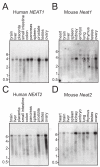
Northern Analysis of NEAT1 and NEAT2/MALAT-1 in mouse and human tissues. Radiolabeled probes to NEAT1 and NEAT2/MALAT-1 show a ~3.5 kb transcript for the human and mouse NEAT1 loci (A,C) and a transcript of more than 6 kb for the human and mouse NEAT2 loci (B,D). The analyses were carried out using First Choice Northern Blots (Ambion), which contain 2 micrograms each of poly(A) RNA from various tissues. Equal loading of RNA is ensured by Ambion for each lot of blots with either GAPDH or beta-actin probes for human or mouse blots respectively. Difficulties in transferring large transcripts to Northern blots may explain the absence of a larger (>17 kb), NEAT1 transcript (data not shown and [29]).
Figure 4
Figure 4
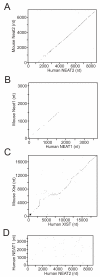
Comparison of NEAT1 and NEAT2/MALAT-1 between the human and mouse genomes. (A) The dot plot details the unspliced 8708 nt of the human NEAT2/MALAT-1 transcript as compared to the 6982 nt mouse Neat2/Malat-1 transcript and shows strong conservation across the length of the NEAT2/MALAT-1 transcript. (B) The dot plot details the 3729 nt human NEAT1 transcript as compared to the 3177 nt mouse Neat1 transcript. Two islands of relatively high identity were identified by this comparison: nucleotide position 483–597 of human NEAT1 and 450–565 of mouse Neat1 show 87% identity, and nucleotide position 1113–1368 of human NEAT1 and 1013–1238 of mouse Neat1 show 80% identity. (C) A dot plot with identical parameters between the primary human and mouse XIST transcripts is shown for comparison. (D) A dot plot between human NEAT1 and NEAT2/MALAT-1 with a 50% identity cutoff demonstrates the lack of conservation between the transcripts.
Figure 5
Figure 5
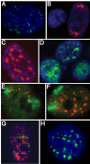
Subcellular localization of human and mouse NEAT1 and NEAT2/MALAT-1. (A) Tig1 cells: Human NEAT1 RNA (green) distributes in discrete foci through the nucleus and is not detected in the cytoplasm. (B) Tig1 cells: NEAT1 RNA (red) is transcribed from 2 loci early in G1 (left) and occasionally shows a highly polarized pattern (right). (C) Tig1 cells: NEAT2/MALAT-1 RNA (red) is not detected in the cytoplasm at interphase and concentrates in domains in the nucleus with weaker signal apparent throughout the nucleoplasm. (D) NIH-3T3s: Mouse Neat2/Malat-1 RNA (green) localizes as bright nucleoplasmic signal in some cells and concentrates in domains in others. (E) NEAT1 RNA (red) does not localize to its parent chromosome 11 (green). (F) NEAT2/MALAT-1 RNA (red) rapidly disperses from its site of transcription (Chromosome 11 (green)) and distributes throughout the nucleus. (G) Tig1 cells: NEAT1 RNA (green) and NEAT2/MALAT-1 RNA are associated in the interphase nucleus. (H) MEFs: Mouse Neat1 RNA (red) is exclusively nuclear, and is found in fewer, larger foci than in human cells, yet shows the same peripheral association with Neat2/Malat-1 RNA (green), and neither RNA associates significantly with DAPI dense heterochromatin (blue).
Figure 6
Figure 6
Human and mouse NEAT1 and NEAT2/MALAT-1 are intimately associated with SC35 domains. (A) Tig1 cells: NEAT2/MALAT-1 RNA (red) is found in every SC35 domain (green); (B) NIH-3T3s: Neat2/Malat-1 RNA (green) concentrates in domains in about 50% of the population, when it does, it overlaps with SRM-300 speckles (red); (C,D,E) NEAT2/MALAT-1 RNA (green (C)) and a probe to poly(A) (red (D)) overlap (yellow (E)) in domains in the nucleus; (F) Neat1 RNA (red) preferentially localizes to the periphery of SRM-300 domains (green) in mouse cells; (G) NEAT1 RNA (red) preferentially associates at the periphery of SC35 domains (green) in human cells; (H) Two transcription sites of NEAT2/MALAT-1 RNA (red) localize with SC35 domains (green) in early G1 daughter cells; (I) Poly(A) domains (red) are apparently formed prior6=- to the spread of NEAT2/MALAT-1 RNA (green) in early G1.

References

    1. Mattick JS, Makunin IV. Non-coding RNA. Hum Mol Genet. 2006;15 Spec No 1:R17–29. doi: 10.1093/hmg/ddl046. - DOI - PubMed
    1. Morey C, Avner P. Employment opportunities for non-coding RNAs. FEBS Lett. 2004;567:27–34. doi: 10.1016/j.febslet.2004.03.117. - DOI - PubMed
    1. Kelley RL, Kuroda MI. The role of chromosomal RNAs in marking the X for dosage compensation. Curr Opin Genet Dev. 2000;10:555–561. doi: 10.1016/S0959-437X(00)00127-1. - DOI - PubMed
    1. Avner P, Heard E. X-chromosome inactivation: counting, choice and initiation. Nat Rev Genet. 2001;2:59–67. doi: 10.1038/35047580. - DOI - PubMed
    1. Clemson CM, McNeil JA, Willard HF, Lawrence JB. XIST RNA paints the inactive X chromosome at interphase: evidence for a novel RNA involved in nuclear/chromosome structure. J Cell Biol. 1996;132:259–275. doi: 10.1083/jcb.132.3.259. - DOI - PMC - PubMed

Publication types

LinkOut - more resources