

Staphylococcus aureus protein A induced inflammatory response in human corneal epithelial cells
- PMID: 17270147
- PMCID: PMC1864947
- DOI: 10.1016/j.bbrc.2007.01.072
Staphylococcus aureus protein A induced inflammatory response in human corneal epithelial cells
Abstract
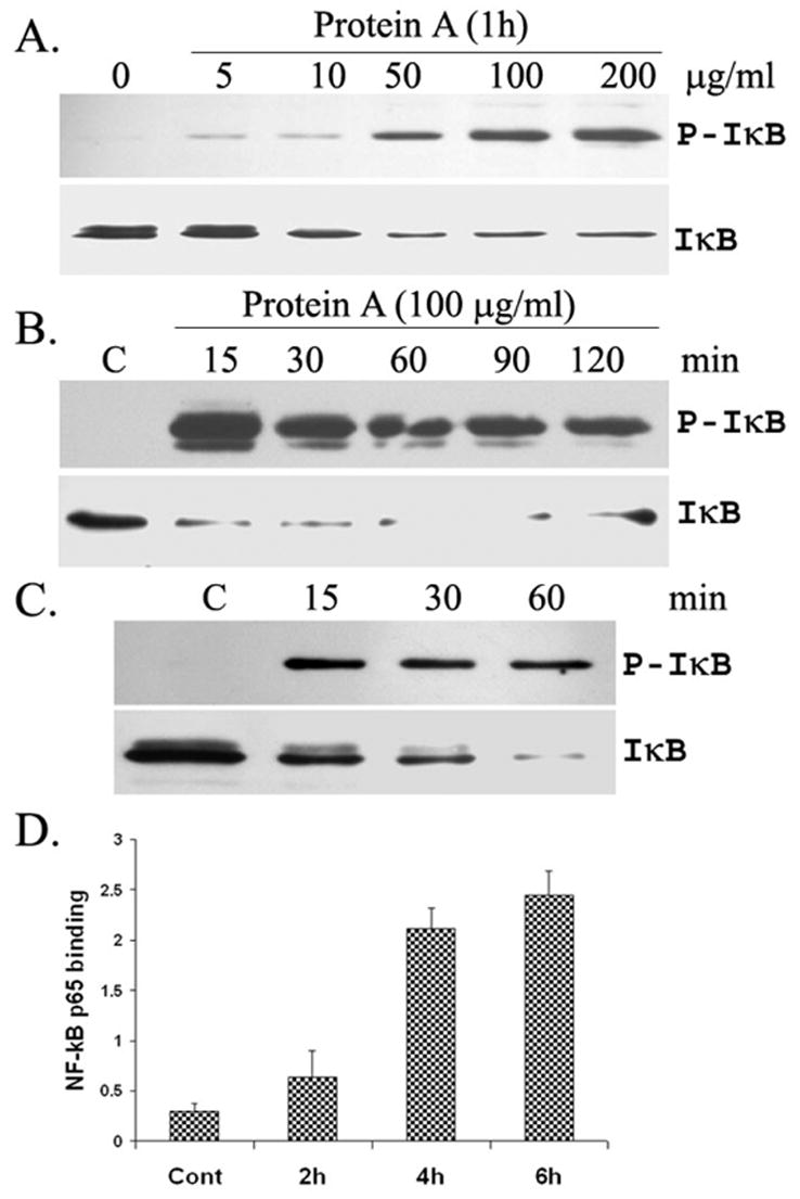
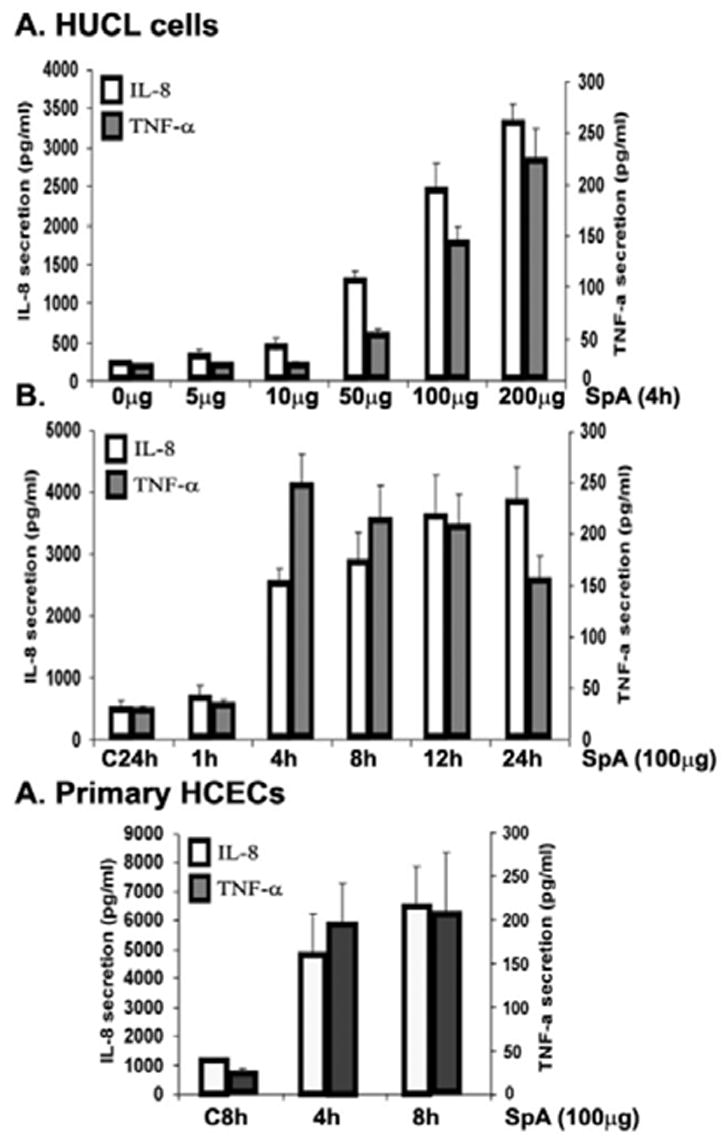
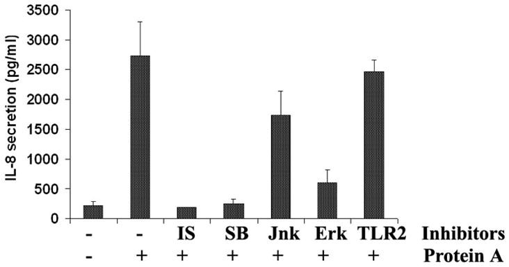
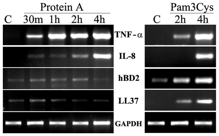
In the present study, we examined the role of Staphylococcus aureus protein A (SpA) in inducing inflammatory response in human corneal epithelial cells (HCECs). Exposure of HCECs to SpA induces rapid NF-kappaB activation and secretion of proinflammatory cytokine/chemokines (TNF-alpha and IL-8) in both concentration and time-dependent manner. Challenge of HCECs with live SpA(-/-) mutant S. aureus strains resulted in significantly reduced production of the cytokines when compared to the wild-type S. aureus strain. SpA also elicited the activation of MAP Kinases P38, ERK, but not JNK, in HCECs. SpA-induced production of proinflammatory cytokine were completely blocked by the NF-kappaB and p38 inhibitors and partially inhibited by the Jnk inhibitor. Pretreatment with anti-TLR2 neutralizing antibody had no effect on SpA-induced inflammatory response in HCECs, suggesting that this response is independent of TLR2 signaling. Moreover, unlike TLR2 ligands, SpA failed to induce the expression of antimicrobial peptides (hBD2 and LL-37) in HCECs. These studies indicate that SpA is a S. aureus virulence factor that stimulates HCEC inflammatory response through a pathway distinct from TLR2 in HCECs.
Figures
Similar articles
-
Staphylococcus aureus lipoproteins trigger human corneal epithelial innate response through toll-like receptor-2.Microb Pathog. 2008 May;44(5):426-34. doi: 10.1016/j.micpath.2007.11.006. Epub 2007 Nov 28. Microb Pathog. 2008. PMID: 18191935 Free PMC article.
-
Toll-like receptor 2-mediated expression of beta-defensin-2 in human corneal epithelial cells.Microbes Infect. 2006 Feb;8(2):380-9. doi: 10.1016/j.micinf.2005.07.006. Epub 2005 Sep 15. Microbes Infect. 2006. PMID: 16242370 Free PMC article.
-
Triggering of toll-like receptors 2 and 4 by Aspergillus fumigatus conidia in immortalized human corneal epithelial cells to induce inflammatory cytokines.Chin Med J (Engl). 2008 Mar 5;121(5):450-4. Chin Med J (Engl). 2008. PMID: 18364120
-
Innate immune response of corneal epithelial cells to Staphylococcus aureus infection: role of peptidoglycan in stimulating proinflammatory cytokine secretion.Invest Ophthalmol Vis Sci. 2004 Oct;45(10):3513-22. doi: 10.1167/iovs.04-0467. Invest Ophthalmol Vis Sci. 2004. PMID: 15452057 Free PMC article.
-
Modulation of corneal epithelial innate immune response to pseudomonas infection by flagellin pretreatment.Invest Ophthalmol Vis Sci. 2007 Oct;48(10):4664-70. doi: 10.1167/iovs.07-0473. Invest Ophthalmol Vis Sci. 2007. PMID: 17898290 Free PMC article.
Cited by
-
Pattern recognition receptors in microbial keratitis.Eye (Lond). 2015 Nov;29(11):1399-415. doi: 10.1038/eye.2015.118. Epub 2015 Jul 10. Eye (Lond). 2015. PMID: 26160532 Free PMC article. Review.
-
Response of corneal epithelial cells to Staphylococcus aureus.Virulence. 2010 Jul-Aug;1(4):223-35. doi: 10.4161/viru.1.4.11466. Virulence. 2010. PMID: 21178448 Free PMC article.
-
Flagellin suppresses the inflammatory response and enhances bacterial clearance in a murine model of Pseudomonas aeruginosa keratitis.Infect Immun. 2008 Jan;76(1):89-96. doi: 10.1128/IAI.01232-07. Epub 2007 Oct 15. Infect Immun. 2008. PMID: 17938214 Free PMC article.
-
Differential response of bovine mammary epithelial cells to Staphylococcus aureus or Escherichia coli agonists of the innate immune system.Vet Res. 2013 Jun 11;44(1):40. doi: 10.1186/1297-9716-44-40. Vet Res. 2013. PMID: 23758654 Free PMC article.
-
Staphylococcus aureus lipoproteins trigger human corneal epithelial innate response through toll-like receptor-2.Microb Pathog. 2008 May;44(5):426-34. doi: 10.1016/j.micpath.2007.11.006. Epub 2007 Nov 28. Microb Pathog. 2008. PMID: 18191935 Free PMC article.
References
-
- Kurpakus-Wheater M, Kernacki KA, Hazlett LD. Maintaining corneal integrity how the “window” stays clear. Prog Histochem Cytochem. 2001;36:185–259. - PubMed
-
- Silverman GJ, Goodyear CS. Confounding B-cell defences: lessons from a staphylococcal superantigen. 2006;6:465–475. - PubMed
-
- Foster TJ. Immune evasion by staphylococci. 2005;3:948–958. - PubMed
Publication types
MeSH terms
Substances
Grants and funding
LinkOut - more resources
Full Text Sources
Research Materials
Miscellaneous
