

Fluorescent resonance energy transfer: A tool for probing molecular cell-biomaterial interactions in three dimensions
- PMID: 17270268
- PMCID: PMC2176075
- DOI: 10.1016/j.biomaterials.2007.01.023
Fluorescent resonance energy transfer: A tool for probing molecular cell-biomaterial interactions in three dimensions
Abstract
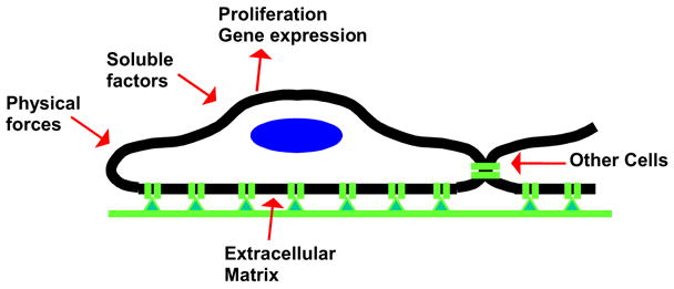
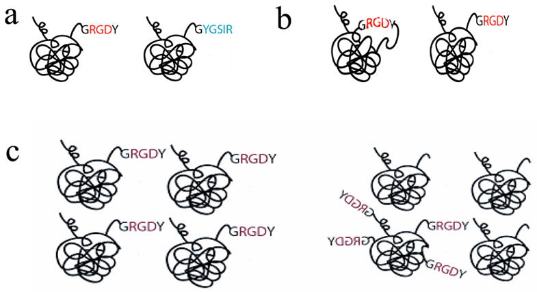
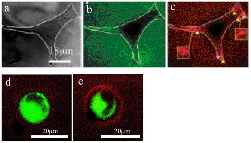
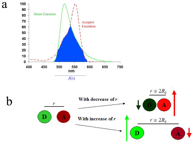
The current paradigm in designing biomaterials is to optimize material chemical and physical parameters based on correlations between these parameters and downstream biological responses, whether in vitro or in vivo. Extensive developments in molecular design of biomaterials have facilitated identification of several biophysical and biochemical variables (e.g. adhesion peptide density, substrate elastic modulus) as being critical to cell response. However, these empirical observations do not indicate whether different parameters elicit cell responses by modulating redundant variables of the cell-material interface (e.g. number of cell-material bonds, cell-matrix mechanics). Recently, fluorescence resonance energy transfer (FRET) has been applied to quantitatively analyze parameters of the cell-material interface for both two- and three-dimensional adhesion substrates. Tools based on FRET have been utilized to quantify several parameters of the cell-material interface relevant to cell response, including molecular changes in matrix proteins induced by interactions both with surfaces and cells, the number of bonds between integrins and their adhesion ligands, and changes in the crosslink density of hydrogel synthetic extracellular matrix analogs. As such techniques allow both dynamic and 3-D analyses they will be useful to quantitatively relate downstream cellular responses (e.g. gene expression) to the composition of this interface. Such understanding will allow bioengineers to fully exploit the potential of biomaterials engineered on the molecular scale, by optimizing material chemical and physical properties to a measurable set of interfacial parameters known to elicit a predictable response in a specific cell population. This will facilitate the rational design of complex, multi-functional biomaterials used as model systems for studying diseases or for clinical applications.
Figures
Similar articles
-
Structural changes of fibronectin adsorbed to model surfaces probed by fluorescence resonance energy transfer.J Biomed Mater Res A. 2004 Jun 1;69(3):525-34. doi: 10.1002/jbm.a.30026. J Biomed Mater Res A. 2004. PMID: 15127399
-
An optical method to quantify the density of ligands for cell adhesion receptors in three-dimensional matrices.J R Soc Interface. 2010 Oct 6;7 Suppl 5(Suppl 5):S649-61. doi: 10.1098/rsif.2010.0321.focus. Epub 2010 Jul 29. J R Soc Interface. 2010. PMID: 20671067 Free PMC article.
-
Role of material-driven fibronectin fibrillogenesis in cell differentiation.Biomaterials. 2011 Mar;32(8):2099-105. doi: 10.1016/j.biomaterials.2010.11.057. Epub 2010 Dec 24. Biomaterials. 2011. PMID: 21185593
-
Fabrication methods of an engineered microenvironment for analysis of cell-biomaterial interactions.Biomaterials. 2007 Jan;28(2):126-33. doi: 10.1016/j.biomaterials.2006.08.007. Epub 2006 Aug 30. Biomaterials. 2007. PMID: 16945407 Review.
-
Cell sensing of physical properties at the nanoscale: Mechanisms and control of cell adhesion and phenotype.Acta Biomater. 2016 Jan;30:26-48. doi: 10.1016/j.actbio.2015.11.027. Epub 2015 Nov 17. Acta Biomater. 2016. PMID: 26596568 Review.
Cited by
-
Fluorescent Probes with Förster Resonance Energy Transfer Function for Monitoring the Gelation and Formation of Nanoparticles Based on Chitosan Copolymers.J Funct Biomater. 2023 Jul 27;14(8):401. doi: 10.3390/jfb14080401. J Funct Biomater. 2023. PMID: 37623646 Free PMC article.
-
Gene delivery through the use of a hyaluronate-associated intracellularly degradable crosslinked polyethyleneimine.Biomaterials. 2009 Oct;30(29):5834-43. doi: 10.1016/j.biomaterials.2009.07.012. Epub 2009 Jul 25. Biomaterials. 2009. PMID: 19631979 Free PMC article.
-
3D imaging of tissue integration with porous biomaterials.Biomaterials. 2008 Oct;29(28):3757-61. doi: 10.1016/j.biomaterials.2008.06.018. Epub 2008 Jul 16. Biomaterials. 2008. PMID: 18635260 Free PMC article.
-
Visualization of integrin molecules by fluorescence imaging and techniques.Biocell. 2021;45(2):229-257. doi: 10.32604/biocell.2021.014338. Epub 2021 Feb 19. Biocell. 2021. PMID: 34219865 Free PMC article.
-
Surface biology of collagen scaffold explains blocking of wound contraction and regeneration of skin and peripheral nerves.Biomed Mater. 2015 Dec 23;11(1):014106. doi: 10.1088/1748-6041/11/1/014106. Biomed Mater. 2015. PMID: 26694657 Free PMC article. Review.
References
-
- Alsberg E, Kong HJ, Hirano Y, Smith MK, Albeiruti A, Mooney DJ. Regulating bone formation via controlled scaffold degradation. J Dent Res. 2003;82(11):903–8. - PubMed
-
- Anseth KS, Bowman CN, Brannon-Peppas L. Mechanical properties of hydrogels and their experimental determination. Biomaterials. 1996;17(17):1647–57. - PubMed
-
- Asthagiri AR, Nelson CM, Horwitz AF, Lauffenburger DA. Quantitative relationship among integrin-binding, adhesion and signaling via focal adhesion kinase and extracellular signal-related kinase 2. J Biol Chem. 274(38):27119–27. - PubMed
-
- Augst AD, Kong HJ, Mooney DJ. Alginate Hydrogels as Biomaterials. Macromol Biosci. 2006;6(8):623–33. - PubMed
-
- Bacskai BJ, Skoch J, Hickey GA, Allen R, Hyman BT. Fluorescence resonance energy transfer determinations using multiphoton fluorescence lifetime imaging microscopy to characterize amyloid-beta plaques. J Biomed Opt. 2003;8(3):368–75. - PubMed
Publication types
MeSH terms
Substances
Grants and funding
LinkOut - more resources
Full Text Sources
Other Literature Sources
Research Materials
