Clinico-pathological features and somatic gene alterations in refractory ceramic fibre-induced murine mesothelioma reveal mineral fibre-induced mesothelioma identities
- PMID: 17272307
- PMCID: PMC4749665
- DOI: 10.1093/carcin/bgm023
Clinico-pathological features and somatic gene alterations in refractory ceramic fibre-induced murine mesothelioma reveal mineral fibre-induced mesothelioma identities
Abstract
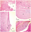
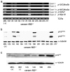
Although human malignant mesothelioma (HMM) is mainly caused by asbestos exposure, refractory ceramic fibres (RCFs) have been classified as possibly carcinogenic to humans on the basis of their biological effects in rodents' lung and pleura and in cultured cells. Hence, further investigations are needed to clarify the mechanism of fibre-induced carcinogenicity and to prevent use of harmful particles. In a previous study, mesotheliomas were found in hemizygous Nf2 (Nf2(+/-)) mice exposed to asbestos fibres, and showed similar alterations in genes at the Ink4 locus and in Trp53 as described in HMM. Here we found that Nf2(+/-) mice developed mesotheliomas after intra-peritoneal inoculation of a RCF sample (RCF1). Clinical features in exposed mice were similar to those observed in HMM, showing association between ascite and mesothelioma. Early passages of 12 mesothelioma cell cultures from ascites developed in RCF1-exposed Nf2(+/-) mice demonstrated frequent inactivation by deletion of genes at the Ink4 locus, and low rate of Trp53 point and insertion mutations. Nf2 gene was inactivated in all cultures. In most cases, co-inactivation of genes at the Ink4 locus and Nf2 was found and, at a lower rate, of Trp53 and Nf2. These results are the first to identify mutations in RCF-induced mesothelioma. They suggest that nf2 mutation is complementary of p15(Ink4b), p16(Ink4a) and p19(Arf) or p53 mutations and show similar profile of gene alterations resulting from exposure to ceramic or asbestos fibres in Nf2(+/-) mice, also consistent with the one found in HMM. These somatic genetic changes define different pathways of mesothelial cell transformation.
Conflict of interest statement
Figures


Similar articles
-
Similar tumor suppressor gene alteration profiles in asbestos-induced murine and human mesothelioma.Cell Cycle. 2005 Dec;4(12):1862-9. doi: 10.4161/cc.4.12.2300. Epub 2005 Dec 7. Cell Cycle. 2005. PMID: 16319530
-
Inactivation of Bap1 Cooperates with Losses of Nf2 and Cdkn2a to Drive the Development of Pleural Malignant Mesothelioma in Conditional Mouse Models.Cancer Res. 2019 Aug 15;79(16):4113-4123. doi: 10.1158/0008-5472.CAN-18-4093. Epub 2019 May 31. Cancer Res. 2019. PMID: 31151962 Free PMC article.
-
A mouse model recapitulating molecular features of human mesothelioma.Cancer Res. 2005 Sep 15;65(18):8090-5. doi: 10.1158/0008-5472.CAN-05-2312. Cancer Res. 2005. PMID: 16166281
-
Animal models of malignant mesothelioma.Inhal Toxicol. 2006 Nov;18(12):1001-4. doi: 10.1080/08958370600835393. Inhal Toxicol. 2006. PMID: 16920675 Review.
-
Molecular pathogenesis of malignant mesothelioma.Carcinogenesis. 2013 Jul;34(7):1413-9. doi: 10.1093/carcin/bgt166. Epub 2013 May 14. Carcinogenesis. 2013. PMID: 23677068 Review.
Cited by
-
Non-neoplastic and neoplastic pleural endpoints following fiber exposure.J Toxicol Environ Health B Crit Rev. 2011;14(1-4):153-78. doi: 10.1080/10937404.2011.556049. J Toxicol Environ Health B Crit Rev. 2011. PMID: 21534088 Free PMC article. Review.
-
Syntenic relationships between genomic profiles of fiber-induced murine and human malignant mesothelioma.Am J Pathol. 2011 Feb;178(2):881-94. doi: 10.1016/j.ajpath.2010.10.039. Am J Pathol. 2011. PMID: 21281820 Free PMC article.
-
Mesotheliomas in Genetically Engineered Mice Unravel Mechanism of Mesothelial Carcinogenesis.Int J Mol Sci. 2018 Jul 27;19(8):2191. doi: 10.3390/ijms19082191. Int J Mol Sci. 2018. PMID: 30060470 Free PMC article. Review.
-
Role of mutagenicity in asbestos fiber-induced carcinogenicity and other diseases.J Toxicol Environ Health B Crit Rev. 2011;14(1-4):179-245. doi: 10.1080/10937404.2011.556051. J Toxicol Environ Health B Crit Rev. 2011. PMID: 21534089 Free PMC article. Review.
-
Molecular Alterations in Malignant Pleural Mesothelioma: A Hope for Effective Treatment by Targeting YAP.Target Oncol. 2022 Jul;17(4):407-431. doi: 10.1007/s11523-022-00900-2. Epub 2022 Jul 30. Target Oncol. 2022. PMID: 35906513 Free PMC article. Review.
References
-
- Pasetto R, et al. Mesothelioma associated with environmental exposures. Med Lav. 2005;96:330–337. - PubMed
-
- Robinson BW, et al. Advances in malignant mesothelioma. N Engl J Med. 2005;353:1591–1603. - PubMed
-
- Mast RW, et al. Studies on the chronic toxicity (inhalation) of four types of refractory ceramic fiber in male fischer 344 rats. Inhal Toxicol. 1995;7:425–467. - PubMed
Publication types
MeSH terms
Substances
LinkOut - more resources
Full Text Sources
Medical
Molecular Biology Databases
Research Materials
Miscellaneous

