. 2007 Apr 15;580(Pt. 2):593-604.
doi: 10.1113/jphysiol.2006.124768. Epub 2007 Feb 1.

Apical GLUT2 and Cav1.3: regulation of rat intestinal glucose and calcium absorption

Affiliations

Apical GLUT2 and Cav1.3: regulation of rat intestinal glucose and calcium absorption

Emma L Morgan et al. J Physiol. .

Abstract

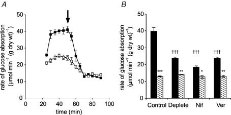
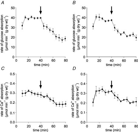
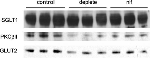
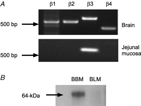
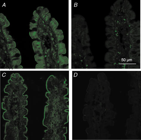
We have proposed a model of intestinal glucose absorption in which transport by SGLT1 induces rapid insertion and activation of GLUT2 in the apical membrane by a PKC betaII-dependent mechanism. Since PKC betaII requires Ca(2+) and glucose is depolarizing, we have investigated whether glucose absorption is regulated by the entry of dietary Ca(2+) through Ca(v)1.3 in the apical membrane. When rat jejunum was perfused with 75 mM glucose, Ca(2+)-deplete conditions, or perfusion with the L-type antagonists nifedipine and verapamil strongly diminished the phloretin-sensitive apical GLUT2, but not the phloretin-insensitive SGLT1 component of glucose absorption. Western blotting showed that in each case there was a significant decrease in apical GLUT2 level, but no change in SGLT1 level. Inhibition of apical GLUT2 absorption coincided with inhibition of unidirectional (45)Ca(2+) entry by nifedipine and verapamil. At 10 mM luminal Ca(2+), (45)Ca(2+) absorption in the presence of 75 mM glucose was 2- to 3-fold that in the presence of 75 mM mannitol. The glucose-induced component was SGLT1-dependent and nifedipine-sensitive. RT-PCR revealed the presence of Ca(v)beta(3) in jejunal mucosa; Western blotting and immunocytochemistry localized Ca(v)beta(3) to the apical membrane, together with Ca(v)1.3. We conclude that in times of dietary sufficiency Ca(v)1.3 may mediate a significant pathway of glucose-stimulated Ca(2+) entry into the body and that luminal supply of Ca(2+) is necessary for GLUT2-mediated glucose absorption. The integration of glucose and Ca(2+) absorption represents a complex nutrient-sensing system, which allows both absorptive pathways to be regulated rapidly and precisely to match dietary intake.


Figures

Figure 1
Figure 1. The effect of luminal Ca2+ and L-type effectors on glucose absorption
A, a representative time course showing the effect of Ca2+. Rat mid and distal jejunum was pre-perfused in vivo in single-pass mode with modified KHB + 100 mm mannitol containing either 1.25 mm Ca2+ (▪) or no Ca2+ (□, Ca2+-deplete perfusate); after 20 min the perfusate was switched to 75 mm glucose + 25 mm mannitol to achieve an initial steady state (30–50 min) before the addition of 1 mm phloretin at 50 min (arrow) to determine the phloretin-insensitive (SGLT1) steady-state rate (60–90 min). B, the effect of L-type effectors, which were present from the start of perfusions. Total (black bars) and phloretin-insensitive (hatched bars) steady-state rates were obtained for control, Ca2+ deplete, nifedipine (10 μm, Nif) and verapamil (100 μm, Ver) perfusions (n=4, each case). Rates are means ± s.e.m. expressed in μmol min−1 (g dry wt)−1. Student's t test was used to determine statistical significance. †††P < 0.001, unpaired test for the comparison of the initial steady-state rates of the Ca2+ deplete, nifedipine and verapamil to that of the control. ***P < 0.001, **P < 0.01, *P < 0.05, paired test comparing the initial steady-state rate with its phloretin-insensitive rate. The phloretin-insensitive rates are not statistically different from one another.
Figure 2
Figure 2. Inhibition of glucose absorption occurs concomitantly with inhibition of 45Ca2+ absorption by L-type channel antagonists
Jejunum was perfused in vivo with perfusate comprising modified KHB containing 75 mm glucose, 1.25 mm Ca2+ and 45Ca2+ (0.35 kBq ml−1) as a tracer to determine unidirectional lumen-to-mucosa 45Ca2+ absorption. After a control period, the perfusion was switched (arrow) to an experimental period in which perfusate contained an L-type antagonist. Time courses are shown for: (A) the rate of glucose absorption when (C) 45Ca2+ absorption was inhibited by nifedipine (10 μm) and for (B) the rate of glucose absorption when (D) 45Ca2+ absorption was inhibited by verapamil (100 μm). Absorption rates are presented as μmol min−1 (g dry wt)−1 and means ± s.e.m., n=4.
Figure 3
Figure 3. Dependence of GLUT2, PKC βII and SGLTI levels on luminal Ca2+ and nifedipine
Brush-border membrane vesicles were prepared from rat jejunum initially perfused in vivo with 75 mm glucose in modified KHB (1.25 mm Ca2+). At 20 min, perfusates were switched to 75 mm glucose in KHB containing either 1.25 mm Ca2+ (control), 0 mm Ca2+ (Ca2+ deplete), or 1.25 mm Ca2+ containing nifedipine (10 μm). Vesicle protein (20 μg) was then separated by SDS-PAGE (10% gels), transblotted on to PVDF and Western blotted for GLUT2, SGLT1 and PKC βII. For full details, see Methods.
Figure 4
Figure 4. Activation of 45Ca2+ absorption by glucose
A, the rates of 45Ca2+ absorption in the presence of either mannitol or glucose were compared within a single perfusion (paired comparison): rat jejunum was perfused for a control period of 40 min with 75 mm glucose and 10 mm Ca2+ (n=4). After 40 min (arrow), perfusion was continued for an experimental period in which 75 mm mannitol was substituted for 75 mm glucose. B, separate perfusions (unpaired comparison): rat jejunum was perfused with 75 mm glucose in the presence of 10 mm Ca2+ for 40 min only (n=8); separate perfusions were undertaken in which mannitol replaced glucose (n=8). P < 0.001 for comparison of the effects of glucose and mannitol in A and B (†††).
Figure 5
Figure 5. Ca2+ absorption at high Ca2+ concentration also displays L-type characteristics
A and C, jejunum was perfused for a control period of 40 min with 75 mm glucose and 10 mm Ca2+ (n=4). After 40 min (arrow), perfusion was continued for an experimental period with 1 mm phloridzin also present in the perfusate. A and C show the glucose and Ca2+ fluxes, respectively. B and D, jejunum was perfused for a control period of 40 min with 75 mm glucose and 10 mm Ca2+ (n=4). After 40 min (arrow), perfusion was continued for an experimental period with 10 μm nifedipine also present in the perfusate. B and D show the glucose and Ca2+ fluxes, respectively. Rates are presented as μmol min−1 (g dry wt)−1.
Figure 6
Figure 6. Rat jejunal mucosa expresses β3 mRNA and β3 protein is located in the apical membrane
A, RT-PCR was performed for the four known auxiliary Cavβ subunits using primers designed to rat specific sequences and cDNA derived from rat brain and rat jejunal mucosa. B, jejunum was perfused for 30 min with 75 mm glucose; brush-border membrane (BBM) and basolateral membrane (BLM) vesicles were then prepared and Western blotted for β3. The BBM band was eliminated by neutralization of antibody with excess peptide (data not shown).
Figure 7
Figure 7. Immunocytochemical localization of Cav1.3 and β3-subunit in the proximal jejunum
Sections of unperfused jejunum were labelled with a rabbit antibody that recognizes all forms of rat Cav1.3 (A and B) and with a rabbit antibody which specifically recognizes the β3 subunit (C and D). The secondary antibody was FITC-conjugated goat anti-rabbit IgG. The peptide controls were sections treated with antibody to Cav1.3 (B) and β3 (D) that had been pre-absorbed with excess antigenic peptide; non-specific staining in the lamina propria of these sections was seen with the secondary alone. All sections are at 63 × magnification and were taken at the same settings with a Zeiss LSM 510 confocal microscope.

References

    1. Affleck J, Helliwell PA, Kellett GL. Immunocytochemical detection of GLUT2 at the rat intestinal brush-border membrane. J Histochem Cytochem. 2003;51:1567–1574. - PMC - PubMed
    1. Au A, Gupta A, Schembri P, Cheeseman CI. Rapid insertion of GLUT2 into the rat jejunal brush-border membrane promoted by glucagon-like peptide 2. Biochem J. 2002;367:247–254. - PMC - PubMed
    1. Auchere D, Tardivel S, Gounelle JC, Drueke T, Lacour B. Role of transcellular pathway in ileal Ca2+ absorption: stimulation by low-Ca2+ diet. Am J Physiol. 1998;275:G951–G956. - PubMed
    1. Auchere D, Tardivel S, Gounelle JC, Lacour B. Stimulation of ileal transport of calcium by sorbitol in in situ perfused loop in rats. Gastroenterol Clin Biol. 1997;21:960–966. - PubMed
    1. Baba R, Yamami M, Sakuma Y, Fujita M, Fujimoto S. Relationship between glucose transporter and changes in the absorptive system in small intestinal absorptive cells during the weaning process. Med Mol Morph. 2005;38:47–53. - PubMed

Publication types

LinkOut - more resources