. 2007 May 1;580(Pt.3):787-800.
doi: 10.1113/jphysiol.2006.121343. Epub 2007 Feb 1.

Associative pairing enhances action potential back-propagation in radial oblique branches of CA1 pyramidal neurons

Affiliations

Associative pairing enhances action potential back-propagation in radial oblique branches of CA1 pyramidal neurons

Sonia Gasparini et al. J Physiol. .

Abstract

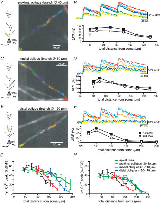
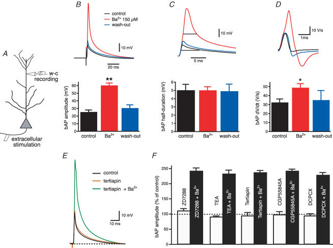
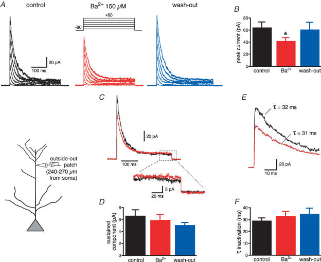
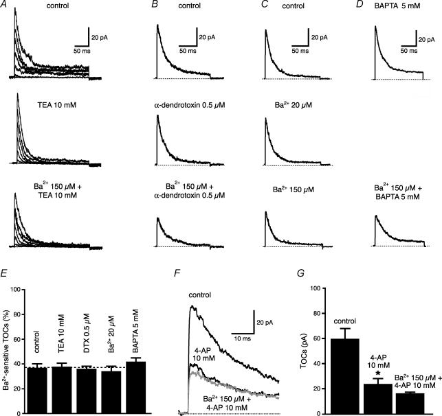
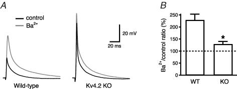
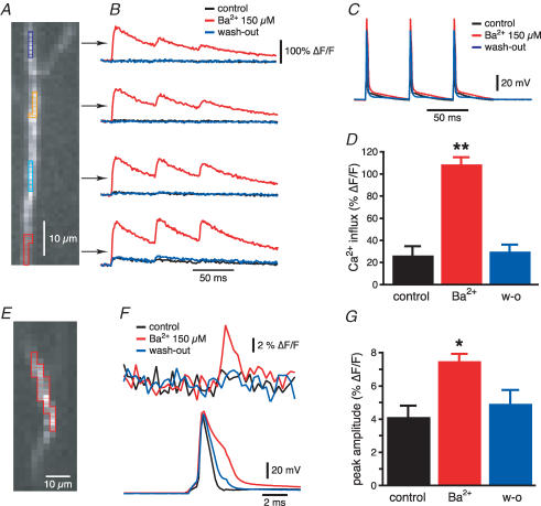
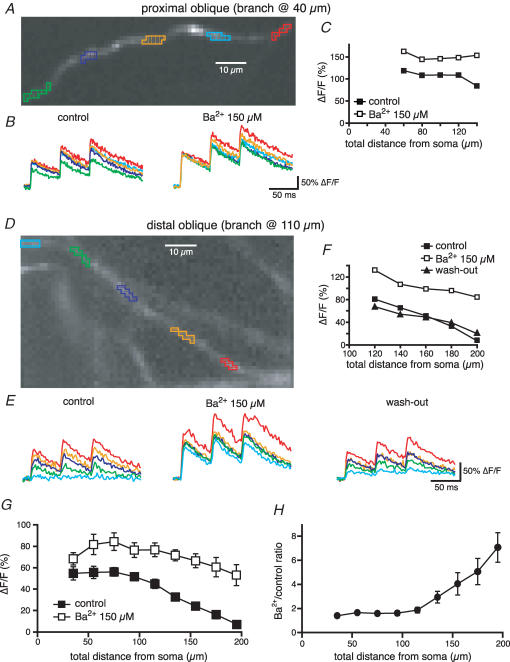
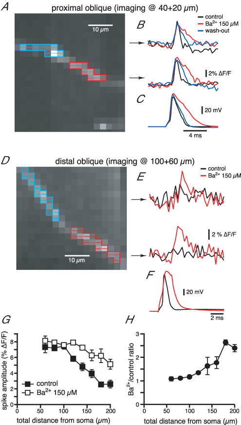
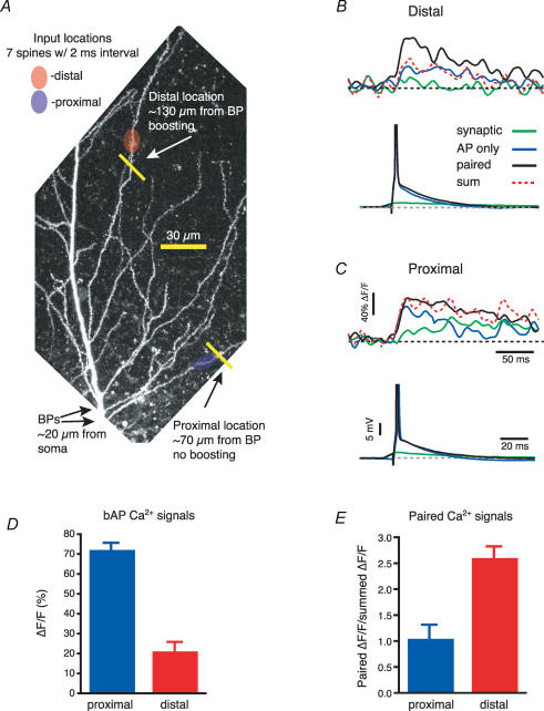
Back-propagating action potentials (bAPs) are involved in associative synaptic plasticity and the modulation of dendritic excitability. We have used high-speed confocal and two-photon imaging to measure calcium and voltage signals associated with action potential propagation into oblique branches of CA1 pyramidal neurons in adult hippocampal slices. The spatial profile of the bAP-associated Ca(2+) influx was biphasic, with an initial increase in the proximity of the branch point followed by a progressive decrease. Voltage imaging in the branches showed that bAP amplitude was initially constant and then steadily declined with distance from the soma. To determine the role of transient K(+) channels in this profile, we used external Ba(2+) (150 microm) as a channel blocker, after characterizing its effect on A-type K(+) channels in the apical trunk. Bath application of Ba(2+) significantly reduced the A-type K(+) current in outside-out patches and nearly eliminated the distance-dependent decrease in bAP amplitude and its associated Ca(2+) signal. Finally, small amplitude bAPs at more distal oblique branch locations could be boosted by simultaneous branch depolarization, such that the paired Ca(2+) signal became nearly the same for proximal and distal oblique dendrites. These data suggest that dendritic K(+) channels regulate the amplitude of bAPs to create a dendritic Ca(2+) signal whose magnitude is inversely related to the electrotonic distance from the soma when bAPs are not associated with a significant amount of localized synaptic input. This distance-dependent Ca(2+) signal from bAPs, however, can be amplified and a strong associative signal is produced once the proper correlation between synaptic activation and AP output is achieved. We hypothesize that these two signals may be involved in the regulation of the expression and activity of dendritic voltage- and ligand-gated ion channels.


Figures

Figure 1
Figure 1. The amplitude of the Ca2+ influx associated with bAP decreases with the distance from the soma in radial oblique branches
A, CCD image of an OGB-1 filled proximal oblique (branch point at 40 μm). B, Ca2+ influxes generated in response to a 20-Hz-train of three somatic action potentials in the regions of the oblique branches marked by like-coloured boxes in A. Below is the plot of the Ca2+ influx associated with the first and the third spike as a function of the distance from the soma. T is the value of ΔF/F recorded at the trunk in the proximity of the branch point. C and D, same as above for a medial oblique (branch point at 80 μm). E and F, same as A and B for a distal oblique (branch point at 130 μm). The insets in A, C and E show the location of the somatic electrode and the imaged region. G, mean calcium influx associated with the first spike of the train as a function of the total distance from the soma. The ΔF/F values for the oblique branches were binned for proximal (branch point between 20 and 60 μm), medial (branch point between 70 and 110 μm) and distal obliques (emerging between 120 and 170 μm). The values of ΔF/F appeared to decline more steeply for the oblique branches than for the apical dendrites for total distances > 120 μm. H, same as G for the third spike.
Figure 2
Figure 2. Ba2+ (150 μm) enhances action potential back-propagation into distal apical dendrites of CA1 pyramidal neurons
A, diagram of experimental configuration for whole-cell recordings from distal apical dendrites (240—270 μm from the soma). B, Ba2+ increases the amplitude of back-propagating action potentials elicited by antidromic stimulation of the axons of CA1 pyramidal neurons, without affecting the half-width (C). D, Ba2+ significantly increased the maximal rate of rise of the bAPs recorded from distal apical dendrites. *P < 0.01, **P < 0.0001. E, the G-protein-activated inward-rectifier K+ (GIRK) channel blocker tertiapin (200 nm) has no effect on bAP amplitude, whereas subsequent application of Ba2+ (150 μm) in the presence of tertiapin enhances back-propagation. F, summary bar graph showing that preincubation with Ih blocker (ZD72288, 20 μm, n = 3), delayed rectifier K+ channel blocker (TEA, 10 mm, n = 3), GIRK channel blocker (tertiapin, 200 nm, n = 6), GABAB receptor antagonist (CGP55845A, 1 μm, n = 2) or adenosine A receptor antagonist (DCPCX, 4 μm, n = 3) could not occlude the effect of Ba2+ (150 μm) on AP back-propagation.
Figure 3
Figure 3. Ba2+-sensitive transient outward currents are expressed in distal apical dendrites of CA1 pyramidal neurons
A, outside-out patches from distal apical dendrites (see inset) were voltage clamped at −80 mV and depolarized to various potentials in the presence of TTX (0.5 μm) to isolate the activity of K+ channels. Ba2+ (150 μm) partially reduced the peak current (see the average plot in B). C, superimposed traces for the step from −80 to +60 mV to show the effect of Ba2+ (red traces) on the transient and the sustained components (shown expanded below). D, mean data show that Ba2+ does not affect the sustained K+ current. E, expanded traces to show the exponential fitting of the decaying phase for the transient K+ current. F, Ba2+ does not affect the time constant of inactivation of the transient K+ current. *P < 0.01
Figure 4
Figure 4. Ba2+-sensitive TOCs are insensitive to various K+ blockers
K+ blockers used: TEA 10 mm for delayed-rectifier (n = 9, A), α-dendrotoxin (500 nm) for Kv1-type (n = 6, B), Ba2+ (20 μm) for inward-rectifier (n = 4, C) and intracellular BAPTA 5 mm for Ca2+-sensitive K+ channel (n = 6, D). E, summary plot of the effect of various K+ channel blockers on TOCs. F, when added to 4-AP (10 mm), Ba2+ has almost no effect, suggesting that they block the same outward current. G, summary plot from F (n = 6).
Figure 5
Figure 5. Deletion of the Kv4.2 gene led to significant reduction of the Ba2+ (200 μm) effect on bAP amplitude in the apical dendrites
A, whole-cell recordings made from approximately 250 μm in apical dendrites of Kv4.2 KO mice and littermate controls. B, the ratio between the bAP amplitude recorded in Ba2+ and in control was significantly (P < 0.01) different for KO mice (128 ± 11%, n = 6) with respect to wild-type littermates (228 ± 23%, n = 6).
Figure 6
Figure 6. Optical imaging data from distal apical dendrites are in agreement with electrophysiological bAP recordings
A, distal portion of the apical dendrite of a CA1 pyramidal neuron filled with Oregon Green BAPTA-1 (100 μm). B, optical recordings of the Ca2+ influx (expressed as ΔF/F) generated in response to the voltage signal in C in the regions of interest marked in A. Ba2+ reversibly increased the amplitude of the Ca2+ signal associated with the bAP. D, average data of the Ba2+-induced increase in the Ca2+ influx in distal apical dendrites at 270 μm from the soma. E, CCD image of a JPW 3028-filled distal apical dendrite. F, voltage signal (above) obtained from the region of interest highlighted in E in response to a somatic action potential (below). G, mean data of the Ba2+-induced increase of the voltage signal in distal apical dendrites at 270 μm. *P < 0.005, **P < 0.0001.
Figure 7
Figure 7. Ba2+ reduces the distance-dependent decrease in the Ca2+ influx associated with bAP in radial oblique branches
A, OGB-1 filled proximal oblique (branch point at 40 μm) from a CA1 pyramidal neuron. B, optical recordings of the Ca2+ influx generated in response to a 20 Hz train of three somatic action potentials in the regions of the oblique branches marked by like-coloured boxes in A in control conditions and in the presence of Ba2+ (150 μm). C, ΔF/F associated with the first spike as a function of the distance from the soma for the oblique branch in A. D, E and F, as above but for a distal radial oblique (branch point at 110 μm). G, mean data for the Ca2+ influx associated with the first bAP as a function of the total distance from the soma in control conditions and in the presence of Ba2+. The effect of Ba2+ is larger for more distal oblique locations, where the Ca2+ influx was reduced in control conditions, as clear from the plot of the ratio in H.
Figure 8
Figure 8. Ba2+ reduces the distance-dependent decrease of the voltage signal associated with bAP in radial obliques
A, CCD image of a JPW 3028-filled proximal radial oblique branch (emerging at 40 μm). B, voltage signals recorded following the somatic action potentials (C) in the two regions highlighted in A in control conditions, in the presence of Ba2+ and upon wash-out. D, E and F, as above for the distal portion of an oblique radial branch. G, mean plot of the voltage signal as a function of the total distance from the soma. As for the Ca2+ influx, the effect of Ba2+ is greater for distal regions of the radial oblique branches, as clear from the plot of the ratio in H.
Figure 9
Figure 9. bAP amplitude in the distal obliques can be boosted by asynchronous synaptic stimulation
A, two photon image stack of the apical trunk and the surrounding oblique branches filled with OGB-1 (100 μm) to show two uncaging and imaging locations. Arrows labelled ‘BPs’ point to the locations where both oblique dendrites branch off from the trunk. B, local Ca2+ transients recorded at the distal location in A for the bAP (blue), the MNI-glutamate asynchronous uncaging (green) and the combination of the two (black). The red dotted line represents the arithmetic sum of the signals obtained during the bAP and the synaptic stimulation. C, as B but for the proximal location. The traces below show the somatic recordings. APs are truncated. D, the plot of the Ca2+ influx for the back-propagating spikes shows a marked decrease with the distance from the soma (proximal = 97 ± 8 μm; distal = 159 ± 4 μm). E, only the distal locations show a significant boosting (expressed as paired ΔF/F/summed ΔF/F) of the bAP by MNI-glutamate uncaging.

References

    1. Alvarez FJ, Dewey DE, Harrington DA, Fyffe RE. Cell-type specific organization of glycine receptor clusters in the mammalian spinal cord. J Comp Neurol. 1997;379:150–170. - PubMed
    1. Andrasfalvy BK, Magee JC. Changes in AMPA receptor currents following LTP induction on rat CA1 pyramidal neurones. J Physiol. 2004;559:543–554. - PMC - PubMed
    1. Andreasen M, Lambert JD. Regenerative properties of pyramidal cell dendrites in area CA1 of the rat hippocampus. J Physiol. 1995;483:421–441. - PMC - PubMed
    1. Antic S, Major G, Zecevic D. Fast optical recordings of membrane potential changes from dendrites of pyramidal neurons. J Neurophysiol. 1999;82:1615–1621. - PubMed
    1. Antic S, Zecevic D. Optical signals from neurons with internally applied voltage-sensitive dyes. J Neurosci. 1995;15:1392–1405. - PMC - PubMed

Publication types

MeSH terms

LinkOut - more resources