. 2007 Feb;117(2):448-56.
doi: 10.1172/JCI29571.

The integrated stress response prevents demyelination by protecting oligodendrocytes against immune-mediated damage

Affiliations

The integrated stress response prevents demyelination by protecting oligodendrocytes against immune-mediated damage

Wensheng Lin et al. J Clin Invest. 2007 Feb.

Abstract

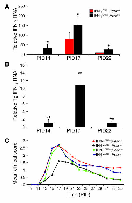
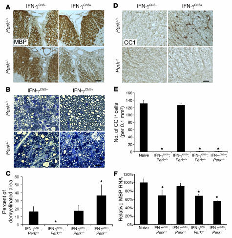
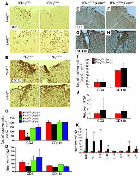
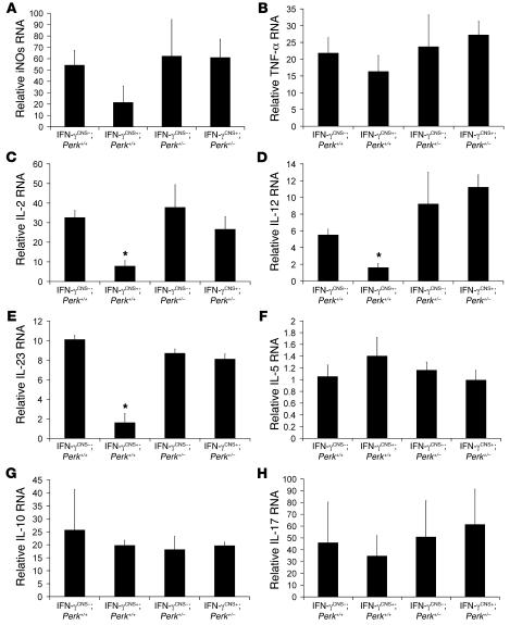
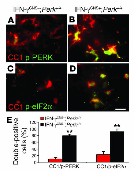
In response to ER stress, the pancreatic endoplasmic reticulum kinase (PERK) coordinates an adaptive program known as the integrated stress response (ISR) by phosphorylating the alpha subunit of eukaryotic translation initiation factor 2 (eIF2alpha). IFN-gamma, which activates the ER stress response in oligodendrocytes, is believed to play a critical role in the immune-mediated CNS disorder multiple sclerosis (MS) and its mouse model, experimental autoimmune encephalomyelitis (EAE). Here we report that CNS delivery of IFN-gamma before EAE onset ameliorated the disease course and prevented demyelination, axonal damage, and oligodendrocyte loss. The beneficial effects of IFN-gamma were accompanied by PERK activation in oligodendrocytes and were abrogated in PERK-deficient animals. Our results indicate that IFN-gamma activation of PERK in mature oligodendrocytes attenuates EAE severity and suggest that therapeutic approaches to activate the ISR could prove beneficial in MS.


Figures

Figure 1
Figure 1. CNS delivery of IFN-γ before EAE onset ameliorated the disease course dependent on the PERK pathway.
(A) Expression pattern of total IFN-γ in the course of EAE (n = 3). (B) Expression pattern of transgenic IFN-γ in the course of EAE (n = 3). (C) Mean clinical score (n = 25). Error bars represent SD. *P < 0.05, **P < 0.01 versus IFN-γCNS–;Perk+/+; Student’s t test.
Figure 2
Figure 2. CNS delivery of IFN-γ before EAE onset protects against EAE-induced demyelination dependent on the PERK pathway.
(A) MBP immunostaining showed that the CNS delivery of IFN-γ protected against EAE-induced demyelination in the lumbar spinal cords of mice on a Perk+/+ background at PID17. In contrast, there was more severe demyelination in the lumbar spinal cord of IFN-γCNS+;Perk+/– mice at PID17 compared with control mice. Scale bar: 50 μm. (B and C) Toluidine blue staining revealed that the myelin and axons in the spinal cords of IFN-γCNS+;Perk+/+ mice remained almost intact at PID17. In contrast, CNS delivery of IFN-γ did not prevent demyelination and axon damage in the lumbar spinal cord of mice on a Perk+/–background at PID17. Scale bar: 10 μm. (D and E) CC1 immunostaining showed that oligodendrocytes in the lumbar spinal cords of IFN-γCNS+;Perk+/+ mice remained almost intact at PID17. In contrast, similar to control mice, IFN-γCNS+;Perk+/– mice lost the majority of oligodendrocytes in the demyelinated lesions of their lumbar spinal cords at PID17. Scale bar: 25 μm. (F) Real-time PCR analysis of the mRNA level of MBP in the indicated spinal cords at PID17 relative to that in the spinal cords of age-matched naive mice. Error bars represent SD. n = 3. *P < 0.05 versus IFN-γCNS–;PERK+/+ (C) or naive mice (E and F).
Figure 3
Figure 3. The effects of IFN-γ on inflammatory infiltration.
(A and C) CD3 immunostaining showed that CNS delivery of IFN-γ reduced T cell infiltration in the lumbar spinal cords of mice on a Perk+/+ background at PID17, but did not significantly affect T cell infiltration in mice on a Perk+/– background. (B and C) CD11b immunostaining revealed that CNS delivery of IFN-γ did not significantly change the numbers of CD11b-positive microglia/macrophages in the lumbar spinal cord of mice on a Perk+/+ or Perk+/– background at PID17 (n = 3). (D) Real-time PCR analysis of the relative mRNA levels of CD3 and CD11b in the spinal cord at PID17 (n = 3). (E, F, and I) CD3 immunostaining showed that CNS delivery of IFN-γ did not affect T cell infiltration in lumbar spinal cord at PID14. (G, H, and I) CD11b immunostaining showed that CNS delivery of IFN-γ did not significantly change the numbers of CD11b-positive microglia/macrophages in the lumbar spinal cord at PID14 (n = 3). (J) Real-time PCR analysis of the relative mRNA levels of CD3 and CD11b in the spinal cord at PID14 (n = 4). (K) Real-time PCR analysis for the expression pattern of cytokines in the spinal cord at PID14 (n = 4). Scale bars: 50 μm. Error bars represent SD. *P < 0.05 versus IFN-γCNS–;Perk+/+.
Figure 4
Figure 4. Real-time PCR analysis for the expression pattern of cytokines in the spinal cord at the peak of disease.
(A) CNS delivery of IFN-γ did not significantly affect the expression of iNOs. (B) CNS delivery of IFN-γ did not significantly affect the expression of TNF-α. (C) CNS delivery of IFN-γ decreased the expression of IL-2 in the spinal cords of mice on a Perk+/+ background, but did not change IL-2 expression in mice on a Perk+/– background. (D) CNS delivery of IFN-γ decreased the expression of IL-12 in the spinal cords of mice on a Perk+/+ background, but did not change IL-12 expression in mice on a Perk+/– background. (E) CNS delivery of IFN-γ decreased the expression of IL-23 in the spinal cords of mice on a Perk+/+ background, but did not change IL-23 expression in mice on a Perk+/– background. (F) CNS delivery of IFN-γ did not significantly affect the expression of IL-5. (G) CNS delivery of IFN-γ did not significantly affect the expression of IL-10. (H) CNS delivery of IFN-γ did not significantly affect the expression of IL-17. Error bars represent SD. n = 3. *P < 0.05 versus IFN-γCNS–;Perk+/+.
Figure 5
Figure 5. The protective effects of IFN-γ in EAE are associated with activation of the PERK-eIF2α pathway in oligodendrocytes.
(A and B) CC1 and p-PERK double labeling showed modest activation of PERK in a few oligodendrocytes in the lumbar spinal cords of control mice at PID14, and CNS delivery of IFN-γ strongly activated PERK in the majority of oligodendrocytes. (C and D) CC1 and p-eIF2α double labeling showed that modest activation of eIF2α in a few oligodendrocytes in the lumbar spinal cord of control mice at PID14, and CNS delivery of IFN-γ markedly activated eIF2α in the majority of oligodendrocytes. Scale bar: 10 μm. (E) Quantitative analysis showed CNS delivery of IFN-γ significantly increased the percentage of double-positive CC1/p-PERK and CC1/p-eIF2α cells in the spinal cords of IFN-γCNS+;Perk+/+ mice compared with IFN-γCNS–;Perk+/+ mice at PID14. Error bars represent SD. n = 3. **P < 0.01 versus IFN-γCNS–;Perk+/+.

Comment in

References

    1. Harding H.P., et al. An integrated stress response regulates amino acid metabolism and resistance to oxidative stress. Mol. Cell. 2003;11:619–633. - PubMed
    1. Proud C.G. eIF2 and the control of cell physiology. Semin. Cell Dev. Biol. 2005;16:3–12. - PubMed
    1. Lu P.D., et al. Cytoprotection by pre-emptive conditional phosphorylation of translation initiation factor 2. EMBO J. 2004;23:169–179. - PMC - PubMed
    1. Buntinx M., Stinissen P., Steels P., Ameloot M., Raus J. Immune-mediated oligodendrocyte injury in multiple sclerosis: molecular mechanisms and therapeutic interventions. Crit. Rev. Immunol. 2002;22:391–424. - PubMed
    1. Frohman E.M., Racke M.K., Raine C.S. Multiple sclerosis—the plaque and its pathogenesis. N. Engl. J. Med. 2006;354:942–955. - PubMed

Publication types

MeSH terms