. 2007 Mar;80(3):485-94.
doi: 10.1086/511888. Epub 2007 Jan 17.

Mutations in cohesin complex members SMC3 and SMC1A cause a mild variant of cornelia de Lange syndrome with predominant mental retardation

Affiliations

Mutations in cohesin complex members SMC3 and SMC1A cause a mild variant of cornelia de Lange syndrome with predominant mental retardation

Matthew A Deardorff et al. Am J Hum Genet. 2007 Mar.

Abstract

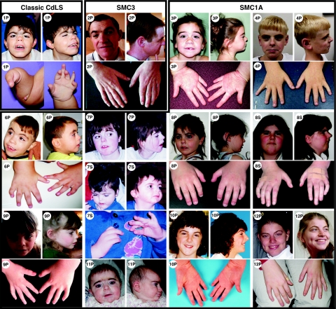
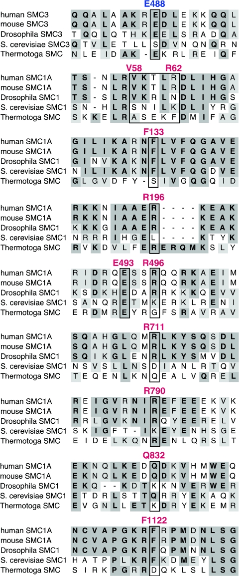
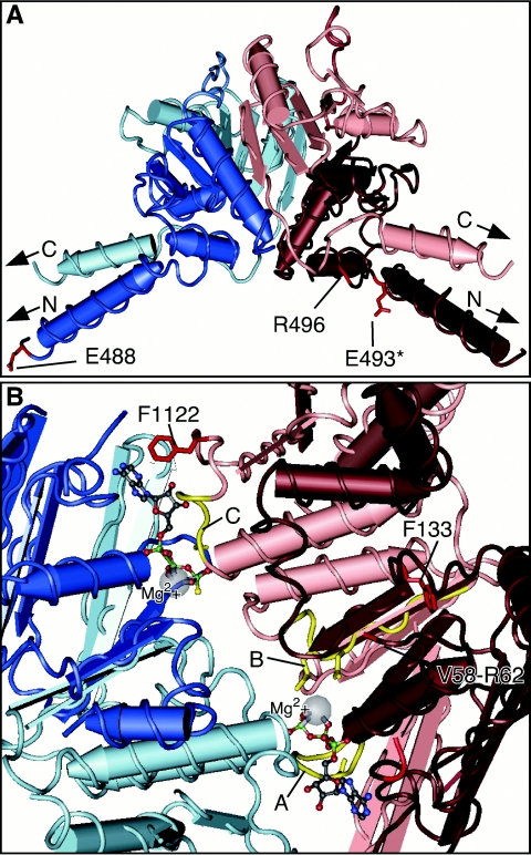
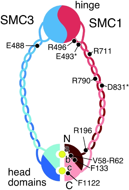
Mutations in the cohesin regulators NIPBL and ESCO2 are causative of the Cornelia de Lange syndrome (CdLS) and Roberts or SC phocomelia syndrome, respectively. Recently, mutations in the cohesin complex structural component SMC1A have been identified in two probands with features of CdLS. Here, we report the identification of a mutation in the gene encoding the complementary subunit of the cohesin heterodimer, SMC3, and 14 additional SMC1A mutations. All mutations are predicted to retain an open reading frame, and no truncating mutations were identified. Structural analysis of the mutant SMC3 and SMC1A proteins indicate that all are likely to produce functional cohesin complexes, but we posit that they may alter their chromosome binding dynamics. Our data indicate that SMC3 and SMC1A mutations (1) contribute to approximately 5% of cases of CdLS, (2) result in a consistently mild phenotype with absence of major structural anomalies typically associated with CdLS, and (3) in some instances, result in a phenotype that approaches that of apparently nonsyndromic mental retardation.


Figures

Figure  1.
Figure 1.
Facies and hands of classic CdLS in SMC3- and SMC1A-mutation–positive individuals. Proband numbers are indicated. A “P” following the number indicates proband, and an “S” indicates an affected sister. Facial features and upper extremities are shown for 1P, a patient with classic CdLS and a truncating NIPBL mutation; 2P, a male with a sporadic SMC3 E488del mutation; 3P, a female with a sporadic V58-R62del mutation; 4P, a male with a sporadic F133V mutation; 6P, a male with a sporadic R496C mutation; 7P and 7S, two sisters of family 2 with the R496H mutation and mosaicism in the unaffected parent; 8P and 8S, two sisters of family 1 who share a R496H mutation; 9P, a female with a sporadic R496H mutation; 10P, a male with a sporadic R711W mutation; 11P, a female with a sporadic R790Q mutation; and 12P, a female with a sporadic F1122L mutation.
Figure  2.
Figure 2.
Conservation of SMC3 and SMC1A and location of mutations. Alignment of human, mouse, Drosophila melanogaster, S. cerevisiae, and T. maritima SMC3- and SMC1-related sequences. Identical residues are indicated in bold with dark shading. Similar residues are indicated in normal font with light shading. Amino acids for which human mutations have been identified are outlined by boxes, with the position indicated above.
Figure  3.
Figure 3.
Mapping of SMC1A and SMC3 mutations to known SMC crystal structure data. A, Mutations in the hinge domain are represented on the Thermotoga SMC dimeric hinge crystal structure. Monomers are colored either red or blue. The darker red and blue regions indicate the N-terminal portions of each monomer. Arrows indicate the N- and C-termini, which are at the end of the coiled-coil domain nearest the hinge region. Mutated residues are shaded bright red. An asterisk (*) indicates the E493 altered residue reported by Musio et al. The R496 residue in humans is a glycine in Thermotoga (see fig. 2). B, Mutations placed on the yeast SMC1 head domain dimer. These regions are key in the binding and hydrolysis of ATP, indicated by ball and stick molecules. Magnesium molecules at the active sites are indicated by gray balls. Three SMC1A mutations occur in the head domain—two in the N-terminus and one in the C-terminus. The V58_R62del overlaps a loop that is unstructured in the yeast SMC1 crystal structure but is positioned near the Walker A and B motifs (yellow residues A and B) and thus could affect ATP binding or hydrolysis. The F133V mutation is adjacent to the Walker B motif that contains a serine, which contacts ATP directly at the active site. The F1122L mutation is in the signature/C motif (yellow residues C) of the C-terminal head domain. It is positioned at the SMC1/SMC3 head-domain intermolecular interface adjoining the adenine ring of ATP. Structural analysis was performed with Cn3D (NCBI Structure Group).
Figure  4.
Figure 4.
Schematic indicating the presumed organization of the SMC1/SMC3 heterodimer and the locations of SMC1A and SMC3 mutations on the cohesin ring. SMC1 is shaded red, and SMC3 is shaded blue, with darker red and blue regions indicating the N-terminal portions of each monomer. Yellow circles indicate ATP molecules bound in proximity to Walker A, B, and C motifs (a, b, and c, respectively). The N-and C-termini are indicated. Mutation locations are indicated by black circles. Altered residues reported by Musio et al. are indicated by an asterisk (*).

References

Web Resources

    1. GenBank, http://www.ncbi.nlm.nih.gov/Genbank/ (for human SMC3 [accession number NM_005445] and human SMC1A [accession number NM_006306])
    1. HUGO Gene Nomenclature Committee, http://www.gene.ucl.ac.uk/nomenclature/
    1. Human Genome Variation Society Mutation Nomenclature Recommendations, http://www.hgvs.org/mutnomen/
    1. NCBI Protein Database, http://www.ncbi.nlm.nih.gov/ (for struture coordinates for the Thermotoga SMC hinge domain [accession numbers 1GXLB and 1GXLA] and S. cerevisiae Smc1 head domain [accession numbers 1W1WA and 1W1WB])
    1. Online Mendelian Inheritance in Man (OMIM), http://www.ncbi.nlm.nih.gov/Omim/ (for CdLS) - PubMed

References

    1. Nasmyth K, Haering CH (2005) The structure and function of SMC and kleisin complexes. Annu Rev Biochem 74:595–64810.1146/annurev.biochem.74.082803.133219 - DOI - PubMed
    1. Hirano T (2006) At the heart of the chromosome: SMC proteins in action. Nat Rev Mol Cell Biol 7:311–32210.1038/nrm1909 - DOI - PubMed
    1. Gillis LA, McCallum J, Kaur M, DeScipio C, Yaeger D, Mariani A, Kline AD, Li HH, Devoto M, Jackson LG, et al (2004) NIPBL mutational analysis in 120 individuals with Cornelia de Lange syndrome and evaluation of genotype-phenotype correlations. Am J Hum Genet 75:610–623 - PMC - PubMed
    1. Krantz ID, McCallum J, DeScipio C, Kaur M, Gillis LA, Yaeger D, Jukofsky L, Wasserman N, Bottani A, Morris CA, et al (2004) Cornelia de Lange syndrome is caused by mutations in NIPBL, the human homolog of Drosophila melanogaster Nipped-B. Nat Genet 36:631–63510.1038/ng1364 - DOI - PMC - PubMed
    1. Tonkin ET, Wang TJ, Lisgo S, Bamshad MJ, Strachan T (2004) NIPBL, encoding a homolog of fungal Scc2-type sister chromatid cohesion proteins and fly Nipped-B, is mutated in Cornelia de Lange syndrome. Nat Genet 36:636–64110.1038/ng1363 - DOI - PubMed

Publication types

MeSH terms

Substances