. 2007 Dec;28(12):1368-75.
doi: 10.1002/hbm.20352.

Relationship between task-related gamma oscillations and BOLD signal: new insights from combined fMRI and intracranial EEG

Affiliations

Relationship between task-related gamma oscillations and BOLD signal: new insights from combined fMRI and intracranial EEG

Jean-Philippe Lachaux et al. Hum Brain Mapp. 2007 Dec.

Abstract

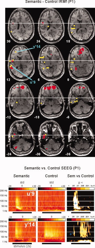
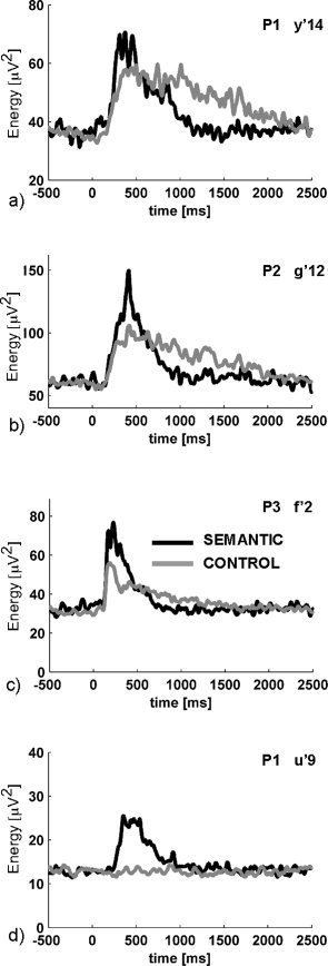
Cognitive neuroscience relies on two sets of techniques to map the neural networks underlying cognition in humans: recordings of either regional metabolic changes (fMRI or PET) or fluctuations in the neural electromagnetic fields (EEG and MEG). Despite major advances in the last few years, an explicit linkage between the two is still missing and the neuroimaging community faces two complementary but unrelated sets of functional descriptions of the human brain. Such an explicit framework, linking the two approaches in potentially complex cognitive tasks and in a variety of brain regions would permit to combine them into fine spatio-temporally-grained human brain mapping procedures. We combined fMRI and intra-cranial EEG recordings of the same epileptic patients during a semantic decision task and found a close spatial correspondence between regions of fMRI activations and recording sites showing EEG energy modulations in the gamma range (>40 Hz). Our findings further support previous findings that gamma band modulations co-localize with BOLD variations and also indicate that fMRI may be used as a constraint to improve source reconstruction of gamma band EEG responses.


Figures

Figure 1
Figure 1
Spatial relationship between BOLD and SEEG gamma modulations. (a) Maximal duration of significant energy difference between the SEMANTIC and the CONTROL conditions in the [40–150 Hz] band, for all the EEG sites. For each site, this value corresponds to the duration of the longest time window, across all frequencies in the gamma band, during which the P‐value for the Mann‐Whitney comparison between the two conditions stays lower than 1 × 10⁻⁴. Sites for which this duration exceeds 100 ms are referred to as “gamma sites” in the text. The markers shapes indicate which patient was recorded; (b) same for the 15 sites for which the maximal duration is longer than 100 ms. Sites closer than 10 mm (resp. 15 mm) away from a fMRI activation cluster (i.e. sets of contiguous voxels above the significance threshold) in the SEMANTIC–CONTROL are shown in green (resp. blue), the remaining sites are shown in red. [Color figure can be viewed in the online issue, which is available at www.interscience.wiley.com.]
Figure 2
Figure 2
Semantic–Control contrasts in fMRI and SEEG (patient P1). The top panel shows the fMRI images of statistically significant increases in BOLD signal from the CONTROL to the SEMANTIC condition (red voxels), thresholded at P < 0.001, superimposed on transverse sections of P1's brain. Yellow dots indicate the brain sites recorded in SEEG, while blue lines point at sites U'9 and Y'14 where task‐related EEG modulations longer than 100 ms were observed in the [40–150 Hz] band. The bottom panels show for each of those sites the time‐frequency maps in the two conditions (left and middle maps, each time‐course is expressed in units of the standard deviation of the [−500:−100 ms] prestimulus period). Time‐frequency maps on the right show the P‐values of a Mann‐Whitney comparison between the SEMANTIC and CONTROL conditions. Regions where the SEMANTIC (resp. CONTROL) condition dominates are indicated by a blue plus (resp. minus) sign. U'9, shows a clear superiority effect of the SEMANTIC condition and is adjacent to a fMRI activation cluster, in the posterior part of the superior temporal gyrus. Y'14, in the inferior frontal gyrus, is >15 mm away from the nearest BOLD cluster, but the effect there is ambiguous: the gamma energy is initially stronger for the semantic condition, before the effect reverses after 500 ms. See the supplementary materials for a similar figure for the other two patients. [Color figure can be viewed in the online issue, which is available at www.interscience.wiley.com.]
Figure 3
Figure 3
Biphasic energy modulations in the gamma band. Each graph shows the mean energy profile in the [40–150 Hz] frequency band for the CONTROL (gray) and SEMANTIC (black) conditions. (a,b,c) correspond to the only sites with a clear condition‐effect in the gamma band which are far (>15 mm) from a BOLD contrast cluster. In all three cases, the initial burst of gamma energy is stronger in the SEMANTIC condition, while its tail is stronger in the CONTROL condition. Such effects, which may be frequent in functional imaging studies, are problematic for fMRI. (d) shows an unambiguous semantic superiority effect for comparison.

References

    1. Aoki F,Fetz EE,Shupe L,Lettich E,Ojemann GA ( 2001): Changes in power and coherence of brain activity in human sensorimotor cortex during performance of visuomotor tasks. Biosystems 63: 89–99. - PubMed
    1. Brovelli A,Lachaux JP,Kahane P,Boussaoud D ( 2005): High gamma frequency oscillatory activity dissociates attention from intention in the human premotor cortex. Neuroimage 28: 154–164. - PubMed
    1. Crone NE,Miglioretti DL,Gordon B,Lesser RP ( 1998a): Functional mapping of human sensorimotor cortex with electrocorticographic spectral analysis. II. Event‐related synchronization in the gamma band. Brain 121 (Part 12): 2301–2315. - PubMed
    1. Crone NE,Miglioretti DL,Gordon B,Sieracki JM,Wilson MT,Uematsu S,Lesser RP ( 1998b): Functional mapping of human sensorimotor cortex with electrocorticographic spectral analysis. I. α and β event‐related desynchronization. Brain 121 (Part 12): 2271–2299. - PubMed
    1. Engel AK,Fries P,Konig P,Brecht M,Singer W ( 1999): Temporal binding, binocular rivalry, and consciousness. Conscious Cogn 8: 128–151. - PubMed

Publication types

MeSH terms