. 2007 Jan-Feb;4(1):119-28.
doi: 10.1021/mp060086o.

Evaluation of an LC8-binding peptide for the attachment of artificial cargo to dynein

Affiliations

Evaluation of an LC8-binding peptide for the attachment of artificial cargo to dynein

Jamie M Bergen et al. Mol Pharm. 2007 Jan-Feb.

Abstract

The limited cytoplasmic mobility of nonviral gene carriers is likely to contribute to their low transfection efficiency. This limitation could be overcome by mimicking the viral strategy of recruiting the dynein motor complex for efficient transport toward the host cell nucleus. A promising approach for attaching artificial cargo to dynein is through an adaptor peptide that binds the 8 kDa light chain (LC8) found in the cargo-binding region of the dynein complex. Several viral proteins that bind LC8 have in common an LC8-binding motif defined by (K/R)XTQT. Short peptides containing this motif have also been shown to bind recombinant LC8 in vitro. However, since the majority of intracellular LC8 exists outside of the dynein complex, it remains unclear whether peptides displaying this LC8-binding motif can access and bind to dynein-associated LC8. In this study, we employed biochemical analysis to investigate the feasibility of attaching artificial cargo to the dynein motor complex using a peptide displaying the well-characterized LC8-binding motif. We report that free intracellular LC8 bound specifically to an LC8-binding (TQT) peptide and not to a control peptide with a mutated LC8-binding motif. However, a similar binding interaction between the TQT peptide and intracellular dynein was not detected. To determine whether dynein binding of the TQT peptide was prevented by competition with free intracellular LC8 or due to the inability of the peptide to access its LC8 binding site in the dynein complex, the TQT peptide was evaluated for its ability to bind either purified LC8 or purified dynein. Our results demonstrate that, while the TQT peptide readily binds free LC8, it cannot bind to dynein-associated LC8. The results emphasize the need to identify functional dynein-binding peptides and highlight the importance of designing peptides that bind to the intact dynein motor complex.


Figures

Figure 1
Figure 1. LC8 from cell lysate specifically binds to the TQT peptide
An affinity pull-down assay was designed to determine whether endogenous LC8 from cell lysate would bind to the immobilized TQT–1 peptide. Peptide-biotin conjugates were immobilized on streptavidin-coated beads and subsequently incubated with HeLa cell lysate. The specificity of this binding interaction was evaluated by immobilizing either a control peptide (CP–1) or biotin alone (no peptide, NP) on the beads. Following elution of bound protein from the beads, LC8 was detected by SDS-PAGE/immunoblot using antibodies against LC8. Cell lysate was used as a positive control for the presence of LC8 (left lane). A sequence-specific interaction between intracellular LC8 and the TQT peptide was evident (TQT–1 lane), while LC8 did not bind to the control peptide or to beads displaying no peptide (CP–1 and NP lanes).
Figure 2
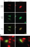
Figure 2. Co-expression of EGFP-LC8 with HcRed-TQT results in co-localization
(A) HeLa cells were co-transfected with plasmids encoding EGFP-LC8 and either HcRed-TQT or HcRed-control peptide (CP). A specific interaction between over-expressed LC8 and the TQT peptide resulted in the formation of punctate intracellular patterns. Co-localization of EGFP-LC8 and HcRed-TQT in these punctate patterns was confirmed using confocal microscopy (left panels). Both LC8 and the control peptide (CP) remained uniformly distributed throughout the cell when co-expressed (right panels). (B) Co-expression of EGFP-LC8 and HcRed-TQT resulted in a variety of intracellular distributions.
Figure 3
Figure 3. The TQT peptide binds to LC8 in cells but not to intact dynein
(A) HeLa cells expressing EGFP-TQT or EGFP-control peptide (CP) were lysed and EGFP was immunoprecipitated using specific antibodies. Co-immunoprecipitated LC8 and dynein intermediate chain (IC74) were detected by SDS-PAGE/immunoblot. Both dynein IC and LC8 were detected in positive control, input lysate samples (Input lanes). LC8, but not dynein IC, co-immunoprecipitated with the TQT peptide (Co-IP, TQT lane), while neither dynein IC nor LC8 co-immunoprecipitated with the control peptide (Co-IP, CP lane). (B) Cells expressing HcRed-TQT or HcRed-CP were lysed and the dynein IC was immunoprecipitated using specific antibodies. LC8 and HcRed were detected by SDSPAGE/immunoblot. As a positive control, both HcRed and dynein LC8 were detectable in the input lysate (Input lanes). LC8 co-immunoprecipitated with the dynein IC in both samples, suggesting the dynein complex remained intact (Co-IP lanes, bottom). However, neither HcRed-TQT nor HcRed-CP co-immunoprecipitated with the dynein complex (Co-IP lanes, top).
Figure 4
Figure 4. The TQT peptide does not co-pellet with dynein and microtubules in a microtubulebinding assay
Interaction between the TQT peptide and the intact dynein complex was examined in the context of a microtubule-binding assay. Purified bovine brain microtubules were incubated with HeLa cell lysate to obtain dynein-enriched microtubule/MAP (microtubule-associated protein) pellets. Streptavidin-biotin-peptide conjugates were incubated with microtubule/MAP samples and subsequently subjected to ultracentrifugation to pellet the microtubules and associated proteins. Supernatant (S) and pellet (P) samples were collected and analyzed by SDS/PAGE followed by either Coomassie blue staining to detect tubulin or immunobloting to detect dynein (IC74) or streptavidinbiotin-peptide conjugates (SA). For both TQT–1 and CP–1 samples, streptavidin-biotin-peptide conjugates remained in the supernatant, while dynein was found in the pellet with the microtubules.
Figure 5
Figure 5. Intact dynein was purified from bovine brain
Cytosol from bovine brain white matter was incubated with taxol to polymerize tubulin into microtubules. The microtubules were then pelleted under ultracentrifugation. Both dynein and kinesin motor proteins pelleted with the microtubules during the first and second wash steps. Incubation with GTP specifically released kinesin from the microtubule pellet. Subsequently, ATP was added to release the dynein complex. Samples of supernatant (S) and pellet (P) were collected at every step. Purification of cytoplasmic dynein from bovine brain was monitored by SDS-PAGE and immunoblot using anti-LC8 antibodies. The dynein heavy chain (HC) partially released from microtubules upon addition of ATP. Co-release of LC8 with the dynein HC suggests that the intact dynein complex was obtained.
Figure 6
Figure 6. Free LC8, but not dynein-associated LC8, binds the immobilized TQT peptide
Equivalent concentrations of either free recombinant LC8 (rLC8) or dynein-associated LC8 were incubated with beads displaying either the TQT or control peptide (CP). Bound proteins were eluted from the beads and LC8 concentration was determined by ELISA. Preferential binding of rLC8 to the TQT-immobilized beads was evident, while dynein-associated LC8 did not bind appreciably to TQT- or CP-immobilized beads. Values presented are the mean fold increase in binding over the control peptide and standard deviation. *, significant statistical difference between rLC8 and dynein groups (p = 0.014).
Figure 7
Figure 7. Model describing the possible TQT/LC8/dynein interactions
LC8 and the TQT peptide can form a binary complex in cells, but cannot form a ternary complex with dynein even in the absence of free competing LC8. (A) The data suggest that free LC8 binds specifically to the TQT peptide and not to a control peptide (CP) lacking the TQT motif. This binding interaction relies on shape complementarity and the formation of specific hydrogen bonds between the TQT motif and residues in the intermonomer groove of LC8., (B) Our results support the inability of an exogenously introduced TQT peptide to bind to dynein-associated LC8. Instead, it is possible that LC8-binding motifs in both intermediate chains (IC) of the dynein motor complex occupy both intermonomer grooves of the LC8 dimer, preventing the attachment cargo to dynein via the TQT-LC8 interactions.

Similar articles

Cited by

References

    1. Lukacs GL, Haggie P, Seksek O, Lechardeur D, Freedman N, Verkman AS. Sizedependent DNA mobility in cytoplasm and nucleus. J Biol Chem. 2000;275:1625–9. - PubMed
    1. Kao HP, Abney JR, Verkman AS. Determinants of the translational mobility of a small solute in cell cytoplasm. J Cell Biol. 1993;120:175–84. - PMC - PubMed
    1. Dowty ME, Williams P, Zhang G, Hagstrom JE, Wolff JA. Plasmid DNA entry into postmitotic nuclei of primary rat myotubes. Proc Natl Acad Sci U S A. 1995;92:4572–6. - PMC - PubMed
    1. Popov S, Poo MM. Diffusional transport of macromolecules in developing nerve processes. J Neurosci. 1992;12:77–85. - PMC - PubMed
    1. Seksek O, Biwersi J, Verkman AS. Translational diffusion of macromolecule-sized solutes in cytoplasm and nucleus. J Cell Biol. 1997;138:131–42. - PMC - PubMed

Publication types