. 2007 Feb;3(2):e18.
doi: 10.1371/journal.ppat.0030018.

Antifungal chemical compounds identified using a C. elegans pathogenicity assay

Affiliations

Antifungal chemical compounds identified using a C. elegans pathogenicity assay

Julia Breger et al. PLoS Pathog. 2007 Feb.

Abstract

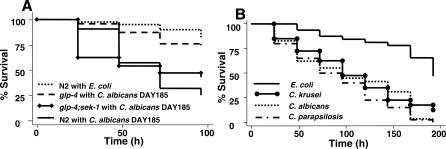
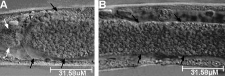
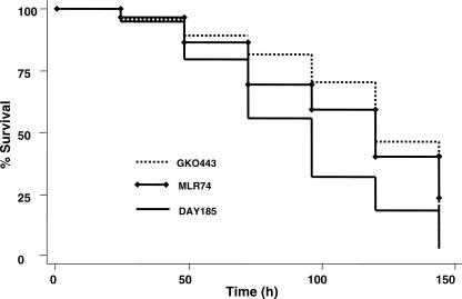
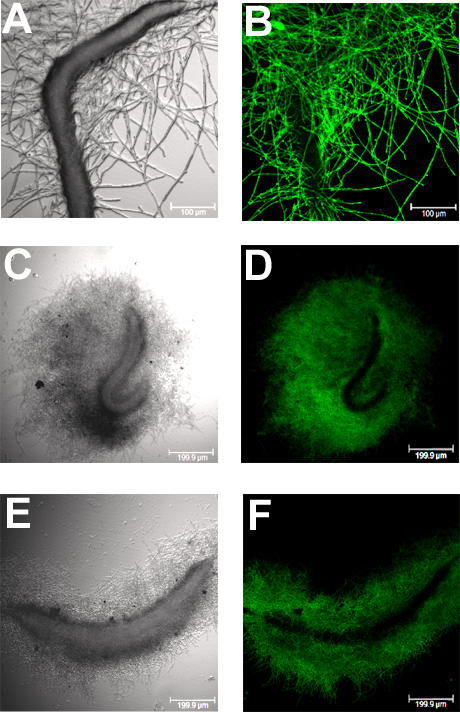
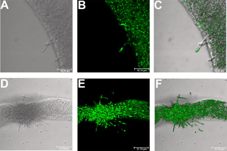
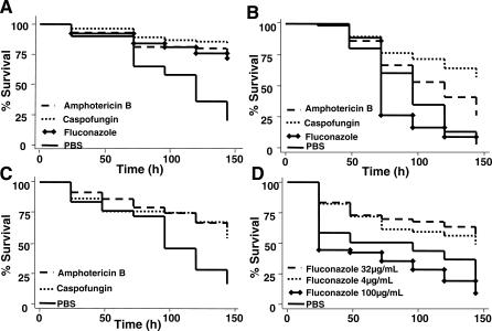
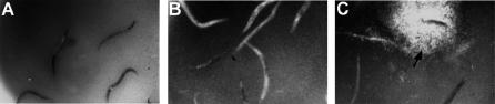
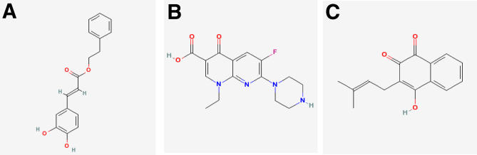
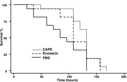
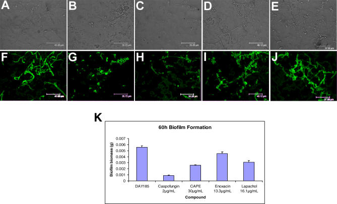
There is an urgent need for the development of new antifungal agents. A facile in vivo model that evaluates libraries of chemical compounds could solve some of the main obstacles in current antifungal discovery. We show that Candida albicans, as well as other Candida species, are ingested by Caenorhabditis elegans and establish a persistent lethal infection in the C. elegans intestinal track. Importantly, key components of Candida pathogenesis in mammals, such as filament formation, are also involved in nematode killing. We devised a Candida-mediated C. elegans assay that allows high-throughput in vivo screening of chemical libraries for antifungal activities, while synchronously screening against toxic compounds. The assay is performed in liquid media using standard 96-well plate technology and allows the study of C. albicans in non-planktonic form. A screen of 1,266 compounds with known pharmaceutical activities identified 15 (approximately 1.2%) that prolonged survival of C. albicans-infected nematodes and inhibited in vivo filamentation of C. albicans. Two compounds identified in the screen, caffeic acid phenethyl ester, a major active component of honeybee propolis, and the fluoroquinolone agent enoxacin exhibited antifungal activity in a murine model of candidiasis. The whole-animal C. elegans assay may help to study the molecular basis of C. albicans pathogenesis and identify antifungal compounds that most likely would not be identified by in vitro screens that target fungal growth. Compounds identified in the screen that affect the virulence of Candida in vivo can potentially be used as "probe compounds" and may have antifungal activity against other fungi.


Conflict of interest statement

Competing interests. The authors have declared that no competing interests exist.

Figures

Figure 1
Figure 1. Killing of C. elegans after Exposure to Candida Species
(A) N2, glp-4, or glp-4;sek-1 L4 nematodes feeding on E. coli OP50 were transferred to C. albicans strain DAY185 for 2 h and then transferred to pathogen-free liquid medium. Dead nematodes were counted and removed daily. N2 worms transferred from E. coli OP50 directly to liquid media were used as control. Survival of glp-4;sek-1 is significantly shorter compared to the strain glp-4 (p < 0.001). (B) Survival of C. elegans glp-4;sek-1 feeding on lawns of C. albicans ATCC#90028, C. parapsilosis ATCC#20019, or C. krusei ATCC#6258. p < 0.001 for each of the yeast strains compared to control nematodes that were exposed to E. coli OP50 (∼65 nematodes/group).
Figure 2
Figure 2. Intact C. albicans Cells Persist within the C. elegans Intestine
C. elegans strain glp-4;sek-1 L4 worms were transferred from OP50 to C. albicans for 30 min and then moved to Candida-free liquid media for 6 d when the worms were photographed. Intact yeast cells are seen within the (A) proximal and (B) distal intestine. White arrows in (A) point to the pharyngeal grinder organ. Black arrows point to the intestinal lumen. Scale bars are 31.58 μm.
Figure 3
Figure 3. Candida Biofilm Formation and Filamentation Is Associated with C. elegans Killing
Survival of C. elegans glp-4;sek-1 animals in liquid pathogen-free media after feeding for 2 h on C. albicans mutants with disruptions in kem1 (strain MLR74) or suv3 (strain GKO443). p = 0.0008 and p < 0.0001, respectively, for the mutants compared to the parental strain C. albicans DAY185 (∼65 nematodes/group).
Figure 4
Figure 4. Progression of Filamentation
The images show C. elegans glp-4;sek-1 nematodes infected by C. albicans strain DAY185 for 2 h and then moved into pathogen-free liquid media. On day 1, dead worms were moved to fresh wells containing liquid media and allowed to incubate until day 6 (A and B), or day 8 (C–F). (A, C, and E) are the light-field images and (B, D, and F) are fluorescent images that show the filaments stained with Concanavalin A-Alexafluor, which binds to polysaccharides. Scale bars are 100 μm (A and B) and 199.9 μm (C–F).
Figure 5
Figure 5. Filamentation Involves the Formation of True Hyphae
Fluorescent microscopy of C. elegans glp-4;sek-1 nematodes after feeding for 2 h on C. albicans strain HGFP3 transformed with pHWP1GFP3 that expresses green fluorescence in a hyphae-specific manner. Worms were transferred to Candida-free liquid media for 2 d (A–C) or 6 d (D–F). Green fluorescence is seen in hyphal cells coming out of the nematode as they penetrate the nematode cuticle. Fluorescence is shown in (B and E). (C and F) show overlap images. Scale bars are 16.16 μm (A–C) and 31.72 μm (D–F).
Figure 6
Figure 6. Antifungals Prolong the Survival of C. elegans glp-4;sek-1 Nematodes Infected by Candida spp. and Evaluation of Antifungals in the C. elegans Assay Allows the Concomitant Evaluation of Toxicity
Nematodes were infected with Candida for 2 h and then moved to pathogen-free liquid media in the presence of antifungals or PBS. Dead worms were counted and removed daily. (A) C. albicans strain MLR6. p < 0.0001 for caspofungin (8 μg/ml), amphotericin B (16 μg/ml), and fluconazole (32 μg/ml). (B) C. krusei strain ATCC#6258. p < 0.0001 for caspofungin (8 μg/ml) and amphotericin B (16 μg/ml). (C) C. parapsilosis ATCC#20019. p < 0.0001 for caspofungin (8 μg/ml), amphotericin B (16 μg/ml), and fluconazole (32 μg/ml). (D) C. parapsilosis ATCC#20019. The p-values are 0.0001 for 4 μg/mL, <0.0001 for 32 μg/mL, 0.01 for 100 μg/mL (∼65 nematodes/group).
Figure 7
Figure 7. Phenotypic Assay Detects Compounds with Antifungal Activity
After exposure to strain C. albicans MLR62, C. elegans glp-4;sek-1 nematodes were pipetted into 96-well plates that contained compounds from chemical libraries. Nematodes exposed to compounds that had antifungal efficacy (in this case, CAPE) had no fluorescence within the intestine (A), whereas nematodes exposed to compounds without antifungal efficacy did not demonstrate any movement (B) and developed filaments outside the nematodes (C). Roughly 25 worms were used per well.
Figure 8
Figure 8. Chemical Compounds Identified Using the C. elegans–Candida Screen Tested in a Murine Model of Hematogenous Candidiasis
(A) CAPE, (B) enoxacin, and (C) lapachol (from http://pubchem.ncbi.nlm.nih.gov).
Figure 9
Figure 9. Compounds Identified in the C. elegans Screen Have Activity in a Murine Model of Hematogenous Candidiasis
Survival of mice after tail vein injection of 1 × 10⁶ blastospores of C. albicans DAY185. Mice that received intravenous injection of CAPE (400 mg/kg once as a loading dose, followed by 200 mg/kg maintenance twice daily) or enoxacin (80 mg/kg loading dose, followed by 40 mg/kg maintenance twice daily) survived significantly longer compared to control mice that received placebo (p = 0.008 and 0.04, respectively). There were 16 mice in each group.
Figure 10
Figure 10. In Vitro Efficacy of CAPE, Enoxacin, and Lapachol in Biofilm Formation
DAY185 formed biofilms on silicone squares in the presence of DMSO in PBS (A and F), caspofungin (B and G), CAPE (C and H), enoxacin (D and I), or lapachol (E and J). Silicone squares were observed for filament formation with a 40× oil emersion objective (A–E) and stained with concanavalin A-Alexa 488 to identify the cell membranes of DAY185 with confocal microscopy (F–J). All three compounds significantly decreased the dry mass of the silicone pads (p = 0.020 for caspofungin, CAPE, and lapachol; and p = 0.029 for enoxacin compared to DMSO in PBS control) (K).

References

    1. Mylonakis E, Aballay A. Worms and flies as genetically tractable animal models to study host-pathogen interactions. Infect Immun. 2005;73:3833–3841. - PMC - PubMed
    1. Ewbank JJ. Tackling both sides of the host-pathogen equation with Caenorhabditis elegans . Microbes Infect. 2002;4:247–256. - PubMed
    1. Slavin M, Fastenau J, Sukarom I, Mavros P, Crowley S, et al. Burden of hospitalization of patients with Candida and Aspergillus infections in Australia. Int J Infect Dis. 2004;8:111–120. - PubMed
    1. Kojic EM, Darouiche RO. Candida infections of medical devices. Clin Microbiol Rev. 2004;17:255–267. - PMC - PubMed
    1. Rentz AM, Halpern MT, Bowden R. The impact of candidemia on length of hospital stay, outcome, and overall cost of illness. Clin Infect Dis. 1998;27:781–788. - PubMed

Publication types