Review
. 2007 Feb;81(2):89-131.
doi: 10.1016/j.pneurobio.2006.12.003. Epub 2007 Jan 5.

N-Acetylaspartate in the CNS: from neurodiagnostics to neurobiology

Affiliations
Review

N-Acetylaspartate in the CNS: from neurodiagnostics to neurobiology

John R Moffett et al. Prog Neurobiol. 2007 Feb.

Abstract

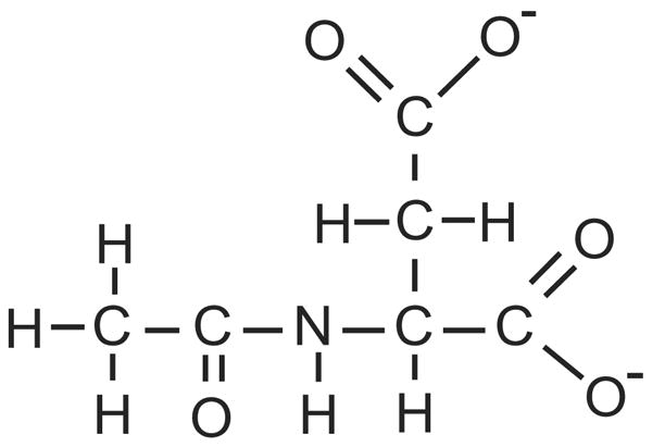
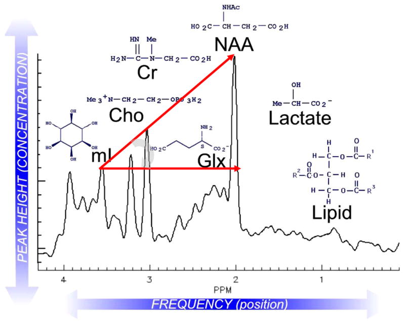
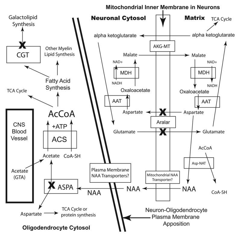
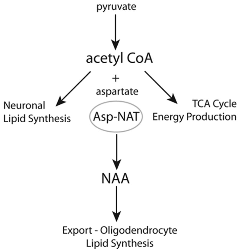
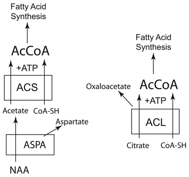
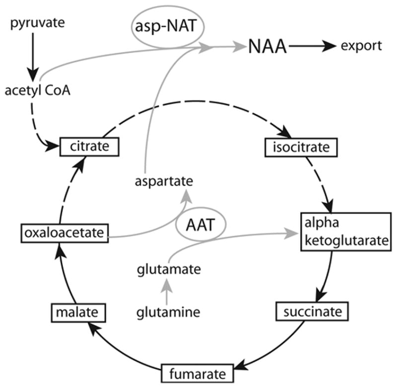
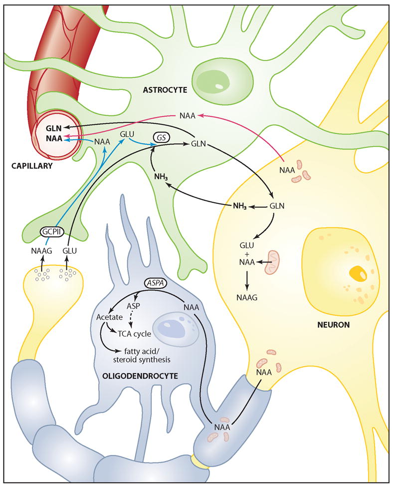
The brain is unique among organs in many respects, including its mechanisms of lipid synthesis and energy production. The nervous system-specific metabolite N-acetylaspartate (NAA), which is synthesized from aspartate and acetyl-coenzyme A in neurons, appears to be a key link in these distinct biochemical features of CNS metabolism. During early postnatal central nervous system (CNS) development, the expression of lipogenic enzymes in oligodendrocytes, including the NAA-degrading enzyme aspartoacylase (ASPA), is increased along with increased NAA production in neurons. NAA is transported from neurons to the cytoplasm of oligodendrocytes, where ASPA cleaves the acetate moiety for use in fatty acid and steroid synthesis. The fatty acids and steroids produced then go on to be used as building blocks for myelin lipid synthesis. Mutations in the gene for ASPA result in the fatal leukodystrophy Canavan disease, for which there is currently no effective treatment. Once postnatal myelination is completed, NAA may continue to be involved in myelin lipid turnover in adults, but it also appears to adopt other roles, including a bioenergetic role in neuronal mitochondria. NAA and ATP metabolism appear to be linked indirectly, whereby acetylation of aspartate may facilitate its removal from neuronal mitochondria, thus favoring conversion of glutamate to alpha ketoglutarate which can enter the tricarboxylic acid cycle for energy production. In its role as a mechanism for enhancing mitochondrial energy production from glutamate, NAA is in a key position to act as a magnetic resonance spectroscopy marker for neuronal health, viability and number. Evidence suggests that NAA is a direct precursor for the enzymatic synthesis of the neuron specific dipeptide N-acetylaspartylglutamate, the most concentrated neuropeptide in the human brain. Other proposed roles for NAA include neuronal osmoregulation and axon-glial signaling. We propose that NAA may also be involved in brain nitrogen balance. Further research will be required to more fully understand the biochemical functions served by NAA in CNS development and activity, and additional functions are likely to be discovered.


Figures

Figure 1
Figure 1
Chemical structure of NAA (mw ~173 Da in the ionic form). The acetate moiety (CH3CO) is on the left, and is attached through the amine nitrogen of aspartate. The 3 roughly equivalent methyl hydrogen atoms on the acetate group resonate with a frequency shift of 2.02 parts per million relative to an MRS standard.
Figure 2
Figure 2
Representative proton MRS spectrum of normal human brain with major peaks of interest depicted. Lactate and lipid signals are absent from this spectrum of a healthy individual. Hunter’s angle (HA; curved gray arrow) refers to the approximate 45-degree angle formed by the peaks myo-inositol (mI), creatine (Cr), choline (Cho), and NAA, when they are present in normal proportions (NAA/Cr ~1.5, Cho/Cr ~0.75; mI/Cr ~0.5) using short-echo-stimulated echo acquisition mode (STEAM) spectroscopy. Changes in HA can be applied to such common MRS diagnoses as tumor (HA < − 50°), stroke, Alzheimer disease (HA ~ 15°), neonatal hypoxia (HA ~ − 45°) or AIDS-related progressive multifocal leukomalacia (HA ~ 0°). Glx = glutamine and glutamate. Reprinted with permission, NeuroRx (Lin et al., 2005).
Figure 3
Figure 3
NAA-immunoreactivity in the rat forebrain. A low magnification photomicrograph of NAA staining in the thalamus and hippocampus is shown in (A). Staining is stronger in most gray matter areas as compared with white matter. Immunoreactivity in the hippocampus is strongest in pyramidal cells, polymorph cells and granule cells (B). Strong NAA-IR is also observed in cortical areas including retrosplenial granular cortex, where both pyramidal and granule cells are strongly immunoreactive (C). In neocortex, staining is particularly strong in layer 5 pyramidal cells, such as those in temporal cortex (D) and motor cortex (E). The columnar organization in these cortical areas can be discerned in NAA-stained sections wherein vertical columns of clustered apical dendrites stained for NAA can be seen (D–F). For methods, see (Moffett and Namboodiri, 1995). Bar = 600 μm A, 100 μm B-E, 50 μm F.
Figure 4
Figure 4
ASPA immunoreactivity in normal and Tremor mutant rat brain and kidney. A section of rat forebrain stained with anti-ASPA antibodies (1:5,000 dilution) is shown in A, whereas a similar section from a Tremor rat ASPA-null mutant is shown stained with the same antibody in B (1:1,000 dilution). In control rat kidney, ASPA expression was strong in proximal tubule cells (C; 1:5,000 dilution), but was not observed in glomeruli (arrows C and D). ASPA immunoreactivity was absent throughout the Tremor rat kidney (D; 1:2,000antibody dilution), Bar = 120 μm.
Figure 5
Figure 5
NAA immunohistochemistry in rat neocortex. NAA is expressed primarily in neurons throughout the brain, but a relatively small number of endothelial cells show strong staining (arrows in A). Stained endothelial cells were observed throughout cortex, including parietal cortex (A and B) and temporal cortex (C and D). Most endothelial cells were unstained for NAA (white holes in tissue, and unstained endothelia in the large vein in A and B). A single endothelial cell in a vein wall is strongly stained for NAA in (B), whereas the other endothelia are unstained. Staining was stronger in endothelial cell nuclei, as opposed to their cytoplasm (arrows C and D). Bar = 60 μm A, 30 μm B, 15 μm C and D.
Figure 6
Figure 6
Simplified schematic of NAA metabolism in neurons and oligodendrocytes, and the relationship to the malate-aspartate mitochondrial shuttle in neurons. The inner mitochondrial membrane in neurons is shown on the right, and the cytoplasm of oligodendrocytes on the left. Genetic mutations or deletions of the genes for the proteins designated by an (X) interrupt the flow of acetate groups from neurons to oligodendrocytes, preventing proper myelin lipid formation. The loss of aspartate-malate shuttle activity in aralar (−/−) mice blocks the ability of neurons to supply NAA to oligodendrocytes, thus compromising myelin lipid synthesis. NAA must be transported out of neurons and into oligodendrocytes at their areas of contact, so we can surmise that specific transporters are involved in NAA flux between them. The fate of NAA derived aspartate in oligodendrocytes is unknown, but it could enter the TCA cycle as oxaloacetate for energy production. Deactivation of ASPA in oligodendrocytes blocks acetate flux from neurons, in the form of NAA, from getting to the cytosol of oligodendrocytes, where myelin lipid synthesis occurs. Blocking downstream enzymes in galactolipid synthesis also compromises myelin lipid synthesis, and proper myelination in the CNS. Abbreviations: AAT, aspartate aminotransferase (EC 2.6.1.1); AcCoA, acetyl coenzyme A; ACS, acetyl CoA synthetase (EC 6.2.1.1); AKG-MT, alpha ketoglutarate-malate transporter; CGT, UDP-galactose:ceramide galactosyltransferase; MDH, malate dehydrogenase (EC 1.1.1.38).
Figure 7
Figure 7
Three major fates for acetyl CoA produced in neurons. Acetyl CoA can be used in neurons for lipid synthesis via the ATP citrate lyase pathway, or energy production via the TCA cycle. When local energy and lipid synthesis demands have been met, a third route of acetyl CoA utilization present in neurons is the Asp-NAT mediated synthesis of NAA using aspartate as co-substrate, followed by export to oligodendrocytes for further metabolism. Pathologies that impair neuronal energetics would be expected to reduce NAA production as acetyl CoA is diverted to energy production and membrane lipid synthesis.
Figure 8
Figure 8
Two different methods of acetyl CoA synthesis leading to fatty acid synthesis. The ASPA/NAA system may only be critical for acetyl CoA (AcCoA) synthesis in certain cells types, such as oligodendrocytes (left), whereas the ATP citrate lyase system is present in most cell types (right). In cells other than oligodendrocytes, citrate provides the substrate for acetyl CoA synthesis. In oligodendrocytes, the ATP citrate lyase system is active, but in addition, NAA is a major substrate for the increased acetyl CoA synthesis required during postnatal myelination. In order to participate in lipid synthesis, the acetate derived from NAA in the cytoplasm of oligodendrocytes must be converted to acetyl CoA, possibly by the enzyme acetyl CoA synthetase-1. Abbreviations: AcCoA, acetyl coenzyme A; ACL, ATP citrate lyase (EC 2.3.3.8); ACS, acetyl CoA synthetase-1 (EC 6.2.1.1); ASPA, aspartoacylase (EC 3.5.1.15); CoA-SH, coenzyme A.
Figure 9
Figure 9
Schematic of the proposed “mini-TCA cycle” in neuronal mitochondria (Yudkoff et al., 1994), and the connection to NAA synthesis. Gray lines indicate the neuron-specific portion of the truncated TCA cycle, and the link to NAA synthesis, whereas dashed lines indicate the part of the TCA cycle that can be bypassed when oxidizing glutamate. As glutamate is converted to alpha ketoglutarate, the truncated TCA cycle produces excess aspartate, and the aspartate can be removed by acetylation through Asp-NAT (Madhavarao et al., 2005; Madhavarao and Namboodiri, 2006). The figure emphasizes the central role of aspartate aminotransferase (AAT) in the ability of neuronal mitochondria to bypass the slower citrate synthase reaction, and to oxidize glutamate through the truncated portion of the TCA cycle. There is no net cost in acetyl CoA utilization during the synthesis of NAA via Asp-NAT, because citrate production is circumvented. Reduced citrate production in neurons may reduce substrate availability for local lipid synthesis via the citrate lyase reaction (Figure 8), but extra NAA is generated which can then be exported and used for increased galactocerebroside and steroid synthesis in oligodendrocytes.
Figure 10
Figure 10
Hypothetical, simplified 4-cell model of NAA synthesis and metabolism in the brain. NAA is synthesized in neuronal mitochondria (see Figure 5), and can then either be transported to oligodendrocytes for fatty acid synthesis and energy production, or can be used for the synthesis of NAAG in neurons. NAAG is released from neuronal synapses along with other transmitters, such as glutamate, and the extracellular NAAG is hydrolyzed by GCPII on astrocytes, which then take up the breakdown products (blue lines). Glutamate and ammonia in astrocytes are converted to glutamine by glutamine synthase (GS), and the glutamine can be excreted to the circulation as a nitrogen removal system, or it can be transported back to neurons for reuse. When the brain nitrogen load is high, NAA excretion might act as a secondary nitrogen removal system, wherein NAA could hypothetically be released by neurons to the extracellular space, taken up by astrocytes, and then excreted to the circulation (red lines). Glutamate and glutamine cycling between neurons and astrocytes involves the production and detoxification of ammonia. Abbreviations: ASP, aspartate; ASPA, aspartoacylase; GCPII, glutamate carboxypeptidase II; GLN, glutamine; GLU, glutamate; GS, glutamine synthetase, NAA, N-acetylaspartate; NAAG, N-acetylaspartylglutamate; NH3, ammonia.

References

    1. Abbott C, Bustillo J. What have we learned from proton magnetic resonance spectroscopy about schizophrenia? A critical update. Curr Opin Psychiatry. 2006;19:135–139. - PubMed
    1. Adachi M, Schneck L, Cara J, Volk BW. Spongy degeneration of the central nervous system (van Bogaert and Bertrand type; Canavan’s disease) A review. Hum Pathol. 1973;4:331–347. - PubMed
    1. Akimitsu T, Kurisu K, Hanaya R, Iida K, Kiura Y, Arita K, Matsubayashi H, Ishihara K, Kitada K, Serikawa T, Sasa M. Epileptic seizures induced by N-acetyl-L-aspartate in rats: in vivo and in vitro studies. Brain Res. 2000;861:143–150. - PubMed
    1. Amstutz DR, Coons SW, Kerrigan JF, Rekate HL, Heiserman JE. Hypothalamic hamartomas: Correlation of MR imaging and spectroscopic findings with tumor glial content. AJNR Am J Neuroradiol. 2006;27:794–798. - PMC - PubMed
    1. Anderson KJ, Borja MA, Cotman CW, Moffett JR, Namboodiri MA, Neale JH. N-acetylaspartylglutamate identified in the rat retinal ganglion cells and their projections in the brain. Brain Res. 1987;411:172–177. - PubMed

Publication types