Review
. 2007 Apr 14;145(4):1388-96.
doi: 10.1016/j.neuroscience.2006.12.020. Epub 2007 Feb 1.

Xeroderma pigmentosum, trichothiodystrophy and Cockayne syndrome: a complex genotype-phenotype relationship

Affiliations
Review

Xeroderma pigmentosum, trichothiodystrophy and Cockayne syndrome: a complex genotype-phenotype relationship

K H Kraemer et al. Neuroscience. .

Abstract

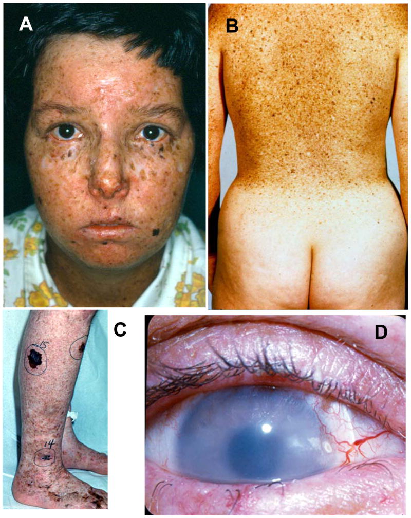
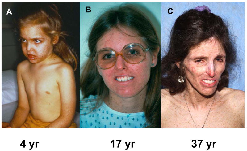
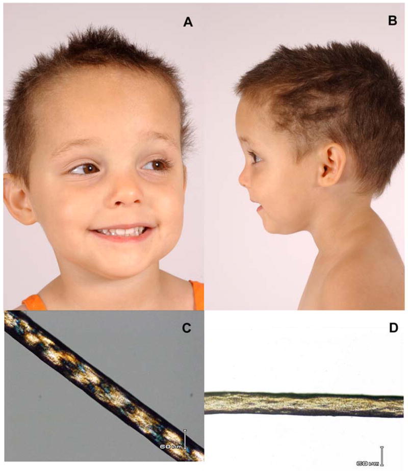
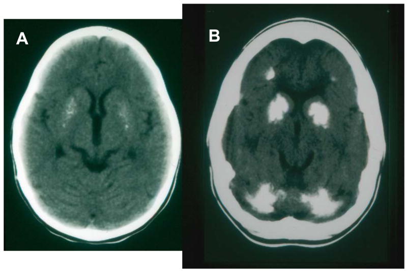
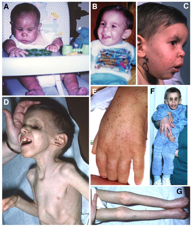
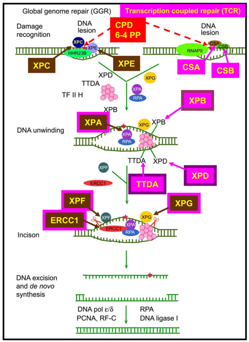
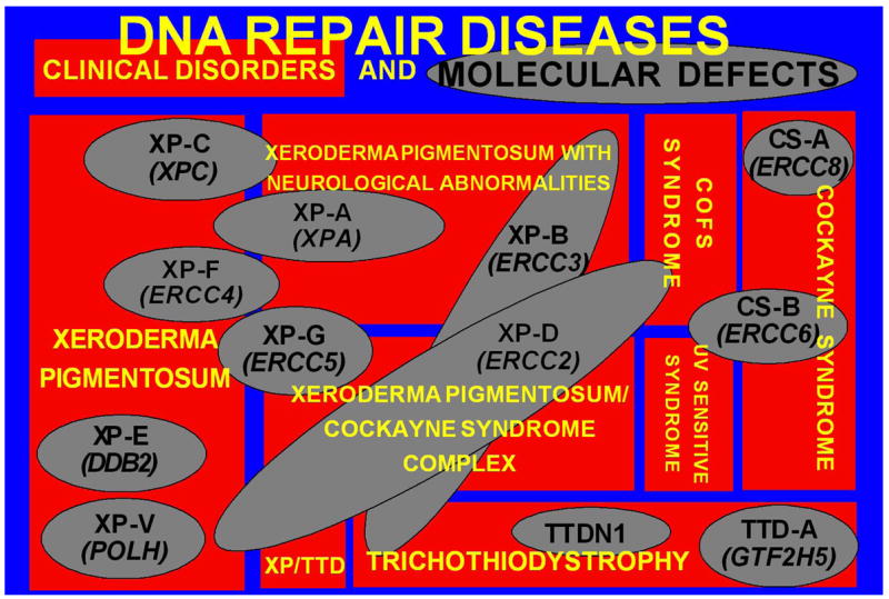
Patients with the rare genetic disorders, xeroderma pigmentosum (XP), trichothiodystrophy (TTD) and Cockayne syndrome (CS) have defects in DNA nucleotide excision repair (NER). The NER pathway involves at least 28 genes. Three NER genes are also part of the basal transcription factor, TFIIH. Mutations in 11 NER genes have been associated with clinical diseases with at least eight overlapping phenotypes. The clinical features of these patients have some similarities but also have marked differences. NER is involved in protection against sunlight-induced DNA damage. While XP patients have 1000-fold increase in susceptibility to skin cancer, TTD and CS patients have normal skin cancer risk. Several of the genes involved in NER also affect somatic growth and development. Some patients have short stature and immature sexual development. TTD patients have sulfur deficient brittle hair. Progressive sensorineural deafness is an early feature of XP and CS. Many of these clinical diseases are associated with developmental delay and progressive neurological degeneration. The main neuropathology of XP is a primary neuronal degeneration. In contrast, CS and TTD patients have reduced myelination of the brain. These complex neurological abnormalities are not related to sunlight exposure but may be caused by developmental defects as well as faulty repair of DNA damage to neuronal cells induced by oxidative metabolism or other endogenous processes.


Figures

Figure 1
Figure 1
Skin and eye involvement in XP. A. Patient XP6BE, 17 year old female with marked poikilodermatous changes of her face with areas of increased freckle-like pigmentation, areas of decreased pigment and atrophy. The lips show cheilitis and there is absence of eye lashes on both lower lids. She had multiple surgeries for removal of skin cancers including basal cell carcinoma and melanoma. B. Back of patient XP5BE, age 26 years, sister of XP6BE. Her skin shows marked poikilodermatous changes with a sharp cutoff at her sun protected buttocks. These sisters had XP neurological disease. They inherited a defect in the XPD (ERCC2) DNA repair gene. Detailed information is reported in . C. Right calf of patient XP1BE at age 46 years. She had multiple large pigmented lesions. Lesion 15 was a large melanoma that measured 0.55 mm in depth when excised. D. Right eye of patient XP1BE at age 29 years. There is clouding of the cornea, prominent vascular growth on the conjunctiva approaching the limbus and loss of lashes on the lower lid. She is wearing a contact lens. She subsequently had a corneal transplant and an ocular squamous cell carcinoma that required enucleation. This patient inherited a defect in the XPC DNA repair gene. Detailed information is reported in ,
Figure 2
Figure 2
Progressive cachexia in patient XP12BE. At age 4 years she had prominent pigmentary changes on the sun exposed portions of her body (face, arms) with sparing of her chest. By age 17 years she had had many basal cell and squamous cell carcinomas excised. Her main neurological abnormalities were loss of deep tendon reflexes and bilateral sensorineural hearing loss. By age 37 years she had become cachectic, was unable to swallow her food and required assistance to walk. Details of her neurological status up to age 22 years are described in . This patient inherited a defect in the XPA DNA repair gene .
Figure 3
Figure 3
Clinical appearance of TTD. A, B) Three year old female with short, brittle hair which is sparse and broken off at different lengths. She rarely has haircuts except to trim uneven areas. C) Tiger-tail appearance of hair with polarizing microscopy. D) Irregular, undulating hair shaft with light microscopy. From
Figure 4
Figure 4
Calcification of brain in CS. A. Axial brain CT scan of a 3 year old girl showing bilateral and symmetric speckled calcifications of the putamen. Tomographic sections at higher levels (not shown) revealed mild enlargement of the lateral ventricles, decreased attenuation in the white matter due to leukodystrophy and thick skull. She had CS with skin sun sensitivity, short stature and progressive neurological degeneration. B. Axial brain CT scan of a 16 year old girl showing extensive bilateral and symmetric calcifications in the putamen, the globus pallidus and the head of the caudate nucleus. Prominent calcifications are also identified in both cerebellar hemispheres. Less prominent calcifications were present at the gray-white matter junction of the frontal and the parietal lobes which were appreciated in higher tomographic sections. Furthermore in the higher sections there was evident leukodystrophy in the white matter of the cerebral hemispheres and increased skull thickness. She had skin sun sensitivity, short statue and progressive neurological degeneration. Modified from .
Figure 5
Figure 5
Patient XP20BE with XP/CS complex. A. Patient at age 4 months after he developed severe sunburn in March, the first sign of illness. Note the sharp change in redness on his lower legs where his socks blocked some sunlight. B. Normal fullness of the face at age 1 year. C. Age 1.5 years. Characteristic skin lesions of XP: freckling and hypopigmented macules of skin and lips. A benign pedunculated lesion is circled. D. Age 6 years. Typical appearance of CS with deep set eyes, prominent ears, and profound cachexia with posturing of the hand. E. Age 6 years. Pigmentary changes and wrinkling of the dorsum of the hand showing signs of premature aging. F. Age 6 years. Signs of advanced CS with profound weakness of dorsiflexion of the hand. Note small size in comparison to the mother who is holding him. G. Profound wasting and contractures of the legs with distal cyanosis and permanently upgoing toes. From
Figure 6
Figure 6
Nucleotide excision repair pathway. Transcription coupled repair (TCR) removes damage from actively transcribing genes while global genome repair (GGR) removes damage from the remainder of the genome. In GGR damage such as ultraviolet induced cyclobutane pyrimidine dimers (CPD) or 6–4 photoproducts (6–4 PP) are recognized by proteins including the XPE (DDB2) and XPC gene products. In TCR, the lesion appears to block the progress of RNA polymerase II in a process involving the CSA and CSB gene products. Following initial damage recognition the pathways converge. The XPB (ERCC3) and XPD (ERCC2) helicases unwind the region surrounding the lesion along with the XPA and XPG (ERCC5) gene products, and replication protein A (RPA). The XPF and XPG (ERCC5) endonucleases perform incisions to remove the lesion in a fragment of about 30 nucleotides. The resulting gap is filled in by de novo DNA synthesis. This system is coordinated so that if one part of the pathway is mutated the entire pathway fails to function normally. Mutations in the genes in rectangles have been associated with clinical disease. This diagram is modified from .
Figure 7
Figure 7
DNA repair diseases - relationship of clinical disorders (red rectangles) to molecular defects (gray ovals) in DNA Repair diseases. Eight clinical diseases are represented. One disease may be caused by mutations in several different genes. Conversely, different mutations in one gene may result in several different clinical diseases. Modified from .

References

    1. Bootsma D, Kraemer KH, Cleaver JE, Hoeijmakers JHJ. Nucleotide excision repair syndromes: xeroderma pigmentosum, Cockayne syndrome, and trichothiodystrophy. In: Vogelstein B, Kinzler KW, editors. The Genetic Basis of Human Cancer. 2. McGraw-Hill; New York: 2002. pp. 211–237.
    1. Brooks PJ, Wise DS, Berry DA, Kosmoski JV, Smerdon MJ, Somers RL, Mackie H, Spoonde AY, Ackerman EJ, Coleman K, Tarone RE, Robbins JH. The oxidative DNA lesion 8,5′-(S)-cyclo-2′-deoxyadenosine is repaired by the nucleotide excision repair pathway and blocks gene expression in mammalian cells. J Biol Chem. 2000;275:22355–22362. - PubMed
    1. Cleaver JE, Thompson LH, Richardson AS, States JC. A summary of mutations in the UV-sensitive disorders: xeroderma pigmentosum, Cockayne syndrome, and trichothiodystrophy. Hum Mutat. 1999;14:9–22. - PubMed
    1. Dollfus H, Porto F, Caussade P, Speeg-Schatz C, Sahel J, Grosshans E, Flament J, Sarasin A. Ocular manifestations in the inherited DNA repair disorders. Surv Ophthalmol. 2003;48:107–122. - PubMed
    1. Dubaele S, Proietti d S, Bienstock RJ, Keriel A, Stefanini M, Van Houten B, Egly JM. Basal transcription defect discriminates between xeroderma pigmentosum and trichothiodystrophy in XPD patients. Mol Cell. 2003;11:1635–1646. - PubMed

Publication types

MeSH terms