. 2007 Feb;12(2):247-60.
doi: 10.1016/j.devcel.2007.01.003.

Chk1 is required for spindle checkpoint function

Affiliations

Chk1 is required for spindle checkpoint function

George Zachos et al. Dev Cell. 2007 Feb.

Abstract

The spindle checkpoint delays anaphase onset in cells with mitotic spindle defects. Here, we show that Chk1, a component of the DNA damage and replication checkpoints, protects vertebrate cells against spontaneous chromosome missegregation and is required to sustain anaphase delay when spindle function is disrupted by taxol, but not when microtubules are completely depolymerized by nocodazole. Spindle checkpoint failure in Chk1-deficient cells correlates with decreased Aurora-B kinase activity and impaired phosphorylation and kinetochore localization of BubR1. Furthermore, Chk1 phosphorylates Aurora-B and enhances its catalytic activity in vitro. We propose that Chk1 augments spindle checkpoint signaling and is required for optimal regulation of Aurora-B and BubR1 when kinetochores produce a weakened signal. In addition, Chk1-deficient cells exhibit increased resistance to taxol. These results suggest a mechanism through which Chk1 could protect against tumorigenesis through its role in spindle checkpoint signaling.


Figures
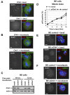
Figure 3
Figure 3. Chk1 Is Required for Localization of BubR1 to Kinetochores during Treatment with Taxol
(A and B) Projected deconvolved image stacks of DT40 and Chk1−/− cells expressing Mad2:GFP and treated with (A) taxol or (B) nocodazole for 45 min. Red, BubR1; green, Mad2:GFP; blue, DNA. The scale bar is 5 μm. (C)Western blot analysis of total Chk1 and actin in BE cells mock transfected (mock), transfected with negative siRNA (control), or transfected with Chk1 siRNA (siChk1). (D)Mitotic index analysis of BE cells transfected as in (C) and treated with taxol. pH3, phosphorylated Ser10 of histone H3. Error bars show the standard deviation from the mean from three experiments. (E and F) Projected deconvolved image stacks of BE cells transfected as in (C) and treated with (E) taxol or (F) nocodazole for 2 hr. Red, BubR1; green, CENP-B; blue, DNA. The scale bar is 5 μm.
Figure 1
Figure 1. Chk1-Deficient Cells Exhibit Chromosome Missegregation, Chromosomal Instability, and Defects in Localization of BubR1 to Kinetochores during Unperturbed Mitosis
(A)Example of chromosome missegregation. A Chk1−/− cell expressing H2B:GFP was analyzed by time-lapse microscopy. Phase-contrast (left panels) and fluorescence images (right panels) of (i) metaphase and (ii) anaphase are shown. (i) Misaligned and (ii) missegregated chromosomes are indicated by arrowheads. The scale bar is 5 μm. (B)Distribution of macrochromosome numbers from mitotic spreads. A total of 60 metaphase spreads were counted for each cell line. (C)BE cells transfected with negative siRNA (control) or Chk1 siRNA (siChk1) were analyzed by confocal microscopy. Examples of missegregated chromosomes during anaphase are indicated by arrowheads. Red, CENP-B; green, α-tubulin; blue, DNA. A single image plane is shown. The scale bar is 5 μm. (D)Projected deconvolved image stacks of DT40 and Chk1−/− cells in prometaphase. Green, BubR1; blue, DNA. The scale bar is 5 μm. (E)Proiected deconvolved image stacks of BE cells transfected as in (C). Red, BubR1; green, CENP-B; blue, DNA. The scale bar is 5 mm. (F)Western blot analysis of BubR1 and actin.
Figure 2
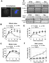
Figure 2. Chk1-Deficient Cells Fail to Sustain Taxol-Induced Mitotic Arrest
(A)Chk1 colocalizes with BubR1 at kinetochores during prometaphase. Example of a Chk1−/−cell expressing Chk1:GFP and analyzed by confocal microscopy. Red, BubR1; green, Chk1:GFP; blue, DNA. A single image plane is shown. The scale bar is 5 μm. (B and C) Mitotic index analysis of G2/M-elutriated DT40, Chk1−/−, and revertant (Rev) cells during treatment with (B) taxol or (C) nocodazole. pH3, phosphorylated Ser10 of histone H3. Error bars show the standard deviation from the mean from three experiments. (D)Upper panels: CDK1-associated histone H1 kinase activity and western blot analysis of CDK1 (Tyr15-phosphorylated and total) and actin during treatment of G2/M-elutriated cells with taxol. Lower panels: western blot analysis of cyclin B2 and actin. Phosphorylated cyclin B2 is marked by an asterisk. (E)Phosphorylated cyclin B2 as a percentage of the total cyclin B2 protein during treatment of G2/M-elutriated cells with taxol. Error bars show the standard deviation from the mean from three experiments. (F)Endoreduplication in Chk1−/− cells during treatment with taxol. Cells were treated as in (B), and the percentage of cells with greater than 4N DNA content was determined. Error bars show the standard deviation from the mean from three experiments.
Figure 4
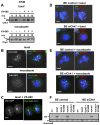
Figure 4. Phosphorylation of Chk1 during Treatment of Cells with Spindle Poisons
(A)Chk1 kinase activity is required for taxol-induced mitotic arrest. Mitotic index analysis of G2/M-elutriated DT40, Chk1−/−, kinase-dead (KD), and revertant (Rev) cells during treatment with taxol. pH3, phosphorylated Ser10 of histone H3. Error bars show the standard deviation from the mean from three experiments. (B)Western blot analysis of total Chk1 and actin in asynchronous cells. (C and D) Western blot analysis of total Chk1 during treatment of G2/M-elutriated DT40 cells with (C) taxol or (D) nocodazole, in the absence or presence of caffeine. The asterisk marks phosphorylated Chk1. Treatment of sample with λ protein phosphatase (PP) is shown. (E)Western blot analysis of total Chk1, phosphorylated Chk1 at Serine 345 (pS345), and actin. G2/M-elutriated DT40 cells were untreated (un) or were treated with taxol or etoposide for 8 hr. (F)Taxol-induced mitotic arrest is weakened by caffeine. Mitotic index analysis of G2/M-elutriated DT40 cells during treatment with taxol, in the presence or absence of caffeine. pH3, phosphorylated Ser10 of histone H3.
Figure 5
Figure 5. Chk1-Deficient Cells Exhibit Decreased Aurora Activity during Treatment with Taxol
(A) Mitotic phosphorylation of Chk1 does not require Aurora activity. Western blot analysis of total Chk1 during treatment of G2/M-elutriated DT40 cells with taxol or nocodazole, in the absence or presence of VX-680. The asterisk marks phosphorylated Chk1. (B and C) Localization of Chk1 to kinetochores is not dependent on Aurora activity. Chk1−/− cells expressing Chk1:GFP were treated with taxol or nocodazole for 2 hr in the (B) absence or (C) presence of VX-680 and were analyzed by confocal microscopy. Red: BubR1; green, Chk1:GFP; blue, DNA. A single image plane is shown. The scale bar is 5μm (D and E) Projected deconvolved image stacks of BE cells transfected with negative siRNA (control) or with Chk1 siRNA(siChk1) and treated with (D) taxol or (E) nocodazole for 2 hr. Red, CENP-A; green, phosphorylated Serine 7 of CENP-A (pS7); blue, DNA. The scale bar is 5 mm. (F) BE cells transfected as in (D) were treated with taxol, nocodazole (nocod), or VX-680 for 6 hr. Upper panel: Aurora-B-associated histone H3 kinase activity. Lower panel: western blot analysis of immunoprecipitated Aurora-B.
Figure 6
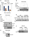
Figure 6. Chk1 Phosphorylates and Enhances Aurora-B Catalytic Activity In Vitro
(A)Treatment with VX-680 exacerbates the checkpoint defect in Chk1−/− cells. Mitotic index analysis of G2/M-elutriated DT40 and Chk1−/− cells during treatment with taxol, in the presence or absence of VX-680. (B)Upper panel: in vitro Chk1 kinase assay with GST, GST-Cdc25C (200-256), and kinase-dead (KD) Aurora-B (D200A) as substrates. Lower panel: western blot analysis of Aurora-B. (C)In vitro Chk1 kinase assay with kinase-active Aurora-B as substrate. (D)Phosphorylation of histone H3 in vitro, with complexes of Aurora-B, Chk1, GST-INCENP (826-919), and kinase-dead (KD) Aurora-B (D200A). pS10, phosphorylated Ser10. Values indicate the relative levels of pS10 of histone H3 with phosphorylation in the presence of Aurora-B arbitrarily set to 1. CBB, Colloidal Brilliant Blue. (E and F) Chk1 is required for taxol-induced phosphorylation of BubR1. (E) Western blot analysis of BubR1 during treatment of G2/M-elutriated cells with taxol/nocodazole, in the presence or absence of VX-680. (F) Western blot analysis of BubR1 in BE cells transfected with negative siRNA (control) orChk1 siRNA (siChk1). Cells were untreated (un) or were treated with taxol/nocodazole (nocod) for 12 hr. The asterisk marks phosphorylated BubR1.
Figure 7
Figure 7. Chk1-Deficient Cells Are Resistant to Killing by Taxol
(A and B) (A) Clonogenic survival and (B) Annexin V staining of DT40 and Chk1−/− cells treated with taxol. Error bars show the standard deviation of the mean from two experiments. un, untreated. (C)Model for the role of Chk1 in the spindle checkpoint.

Comment in

Similar articles

Cited by

References

    1. Biggins S, Murray AW. The budding yeast protein kinase Ipl1/Aurora allows the absence of tension to activate the spindle checkpoint. Genes Dev. 2001;15:3118–3129. - PMC - PubMed
    1. Bishop JD, Schumacher JM. Phosphorylation of the carboxyl terminus of inner centromere protein (INCENP) by the Aurora B Kinase stimulates Aurora B kinase activity. J Biol Chem. 2002;277:27577–27580. - PMC - PubMed
    1. Carvalho A, Carmena M, Sambade C, Earnshaw WC, Wheatley SP. Survivin is required for stable checkpoint activation in taxol-treated HeLa cells. J Cell Sci. 2003;116:2987–2998. - PubMed
    1. Cheeseman IM, Anderson S, Jwa M, Green EM, Kang J, Yates JR, 3rd, Chan CS, Drubin DG, Barnes G. Phospho-regulation of kinetochore-microtubule attachments by the Aurora kinase Ipl1p. Cell. 2002;111:163–172. - PubMed
    1. Collura A, Blaisonneau J, Baldacci G, Francesconi S. The fission yeast Crb2/Chk1 pathway coordinates the DNA damage and spindle checkpoint in response to replication stress induced by topoisomerase I inhibitor. Mol Cell Biol. 2005;25:7889–7899. - PMC - PubMed

Publication types

MeSH terms