

Region-specific mechanisms for testosterone-induced Fos in hamster brain
- PMID: 17276422
- PMCID: PMC1857344
- DOI: 10.1016/j.brainres.2007.01.022
Region-specific mechanisms for testosterone-induced Fos in hamster brain
Abstract
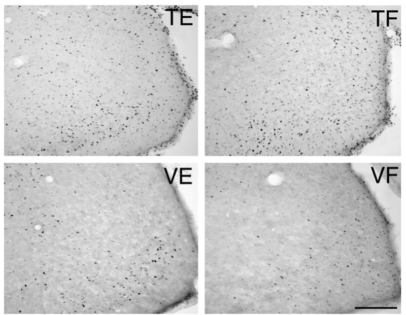
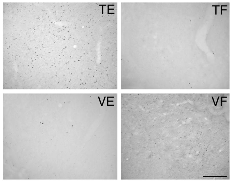
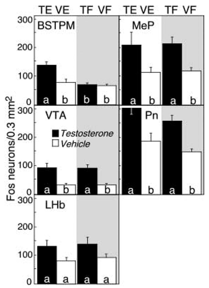
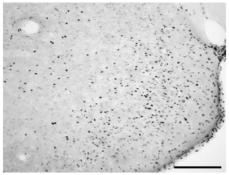
Hamsters self-administer androgens. Previously, we determined that testosterone (T) activates select steroid- and opiate-sensitive brain regions. Is T-stimulated neuronal activation androgenic? Thirty-five castrated males with physiologic T replacement (n=7/group) were pre-treated with the androgen antagonist flutamide (15 mg/kg sc) or ethanol (0.25 ml) and infused into the lateral ventricle (ICV) for 4 h with 40 microg T (TF and TE, respectively) or 40 microl vehicle (VF and VE). To determine if androgens and opiates activate overlapping brain areas, 7 additional males received 20 mug morphine sulfate ICV following ethanol injection (ME). Immediately after ICV infusion, animals were perfused. Sixty-micrometer coronal brain slices were stained for Fos. Fos-positive neurons were counted in a 0.3-mm(2) area from 5 regions previously shown to express T-induced Fos: the posteromedial bed nucleus of the stria terminalis (BSTPM), posteromedial amygdala (MeP), lateral habenula (LHb), ventral tegmental area, and lateral pontine nucleus. T induced Fos in all areas reported previously (TE vs. VE, p<0.05), except LHb (p>0.05). Morphine induced Fos in all 5 brain regions (ME vs. VE, p<0.05), indicating that androgens and opiates activate overlapping brain regions. Flutamide alone did not induce Fos (VF vs. VE, p>0.05). Moreover, flutamide treatment blocked T-induced Fos expression only in the steroid-sensitive BSTPM, suggesting that androgens mediate neuronal activation in this area (mean+/-SEM: TF: 68.4+/-13.2 vs. TE: 137.9+/-17.6, p<0.05). The absence of flutamide effects on T-induced Fos in the steroid-sensitive MeP (TE: 210.6+/-50.0 vs. TF: 215.3+/-28.2, p>0.05) suggests that distinct mechanisms activate Fos in individual androgen-responsive nuclei.
Figures
Similar articles
-
ICV testosterone induces Fos in male Syrian hamster brain.Psychoneuroendocrinology. 2006 Feb;31(2):237-49. doi: 10.1016/j.psyneuen.2005.08.001. Epub 2005 Sep 12. Psychoneuroendocrinology. 2006. PMID: 16157456
-
Study of Fos, androgen receptor and testosterone expression in the sub-regions of medial amygdala, bed nucleus of stria terminalis and medial preoptic area in male Mandarin voles in response to chemosensory stimulation.Behav Brain Res. 2014 Jan 1;258:65-74. doi: 10.1016/j.bbr.2013.10.004. Epub 2013 Oct 12. Behav Brain Res. 2014. PMID: 24129216
-
Androgen dependence in hamsters: overdose, tolerance, and potential opioidergic mechanisms.Neuroscience. 2005;130(4):971-81. doi: 10.1016/j.neuroscience.2004.09.063. Neuroscience. 2005. PMID: 15652994
-
Testosterone differentially influences sex-specific pheromone-stimulated Fos expression in limbic regions of Syrian hamsters.Horm Behav. 1996 Dec;30(4):455-73. doi: 10.1006/hbeh.1996.0050. Horm Behav. 1996. PMID: 9047271
-
Maternal administration of flutamide during late gestation affects the brain and reproductive organs development in the rat male offspring.Neuroscience. 2014 Oct 10;278:122-35. doi: 10.1016/j.neuroscience.2014.07.074. Epub 2014 Aug 15. Neuroscience. 2014. PMID: 25130562
Cited by
-
Gonadal steroid hormone receptors in the medial amygdala contribute to experience-dependent changes in stress vulnerability.Psychoneuroendocrinology. 2021 Jul;129:105249. doi: 10.1016/j.psyneuen.2021.105249. Epub 2021 May 3. Psychoneuroendocrinology. 2021. PMID: 33971475 Free PMC article.
-
Effects of season, testosterone and female exposure on c-fos expression in the preoptic area and amygdala of male green anoles.Brain Res. 2007 Aug 29;1166:124-31. doi: 10.1016/j.brainres.2007.07.004. Epub 2007 Jul 14. Brain Res. 2007. PMID: 17673187 Free PMC article.
-
Aromatase and 5-alpha reductase gene expression: modulation by pain and morphine treatment in male rats.Mol Pain. 2010 Oct 26;6:69. doi: 10.1186/1744-8069-6-69. Mol Pain. 2010. PMID: 20977699 Free PMC article.
-
Effects of testosterone and estradiol on anxiety and depressive-like behavior via a non-genomic pathway.Neurosci Bull. 2015 Jun;31(3):288-96. doi: 10.1007/s12264-014-1510-8. Epub 2015 Mar 9. Neurosci Bull. 2015. PMID: 25754146 Free PMC article.
-
C-fos down-regulation inhibits testosterone-dependent male sexual behavior and the associated learning.Eur J Neurosci. 2013 Nov;38(9):3325-37. doi: 10.1111/ejn.12321. Epub 2013 Jul 29. Eur J Neurosci. 2013. PMID: 23895306 Free PMC article.
References
-
- Amini A, Ahmadiani A. In vivoo evidence for an increase in 5alpha-reductase activity in the rat central nervous system following morphine exposure. Int J Dev Neurosci. 2005;23:621–626. - PubMed
-
- Arvanitogiannis A, Tzschentke TM, Riscaldino L, Wise RA, Shizgal P. Fos expression following self-stimulation of the medial prefrontal cortex. Behav Brain Res. 2000;107:123–132. - PubMed
-
- Aston-Jones G, Harris GC. Brain substrates for increased drug seeking during protracted withdrawal. Neuropharmacology. 2004;47:167–179. - PubMed
-
- Bot G, Chahl LA. Induction of Fos-like immunoreactivity by opioids in guinea-pig brain. Brain Res. 1996;731:45–56. - PubMed
Publication types
MeSH terms
Substances
Grants and funding
LinkOut - more resources
Full Text Sources
Miscellaneous
