. 2007 Apr;53(4):319-31.
doi: 10.1016/j.jinsphys.2006.12.003. Epub 2006 Dec 29.

Seminal proteins but not sperm induce morphological changes in the Drosophila melanogaster female reproductive tract during sperm storage

Affiliations

Seminal proteins but not sperm induce morphological changes in the Drosophila melanogaster female reproductive tract during sperm storage

Erika M Adams et al. J Insect Physiol. 2007 Apr.

Abstract

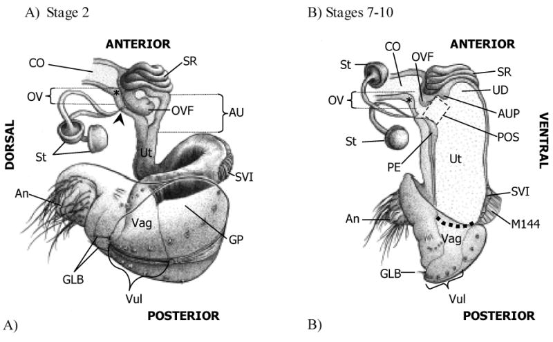
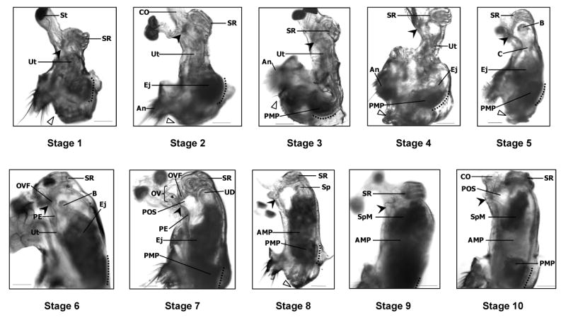
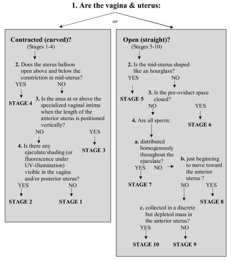
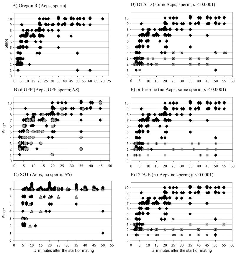
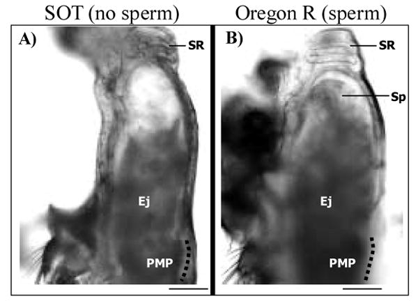
In most insects, sperm transferred by the male to the female during mating are stored within the female reproductive tract for subsequent use in fertilization. In Drosophila melanogaster, male accessory gland proteins (Acps) within the seminal fluid are required for efficient accumulation of sperm in the female's sperm storage organs. To determine the events within the female reproductive tract that occur during sperm storage, and the role that Acps and sperm play in these events, we identified morphological changes that take place during sperm storage in females mated to wild-type, Acp-deficient or sperm-deficient males. A reproducible set of morphological changes occurs in a wild-type mating. These were categorized into 10 stereotypic stages. Sperm are not needed for progression through these stages in females, but receipt of Acps is essential for progression beyond the first few stages of morphological change. Furthermore, females that received small quantities of Acps reached slightly later stages than females that received no Acps. Our results suggest that timely morphological changes in the female reproductive tract, possibly muscular in nature, may be needed for successful sperm storage, and that Acps from the male are needed in order for these changes to occur.


Figures

Figure 1
Figure 1
Conformation of the lower reproductive tract of wild type (Oregon R) Drosophila melanogaster females at early (A) and late (B) times in sperm storage. (A) At the start of ejaculate transfer (Stage 2), the uterus (Ut) is contracted and forms a loop at the specialized vaginal intima (Miller 1950; SVI). In addition, the anterior uterus (AU) is bent laterally above the SVI such that, in the drawing, the SVI and vagina (Vag) appear to project toward the reader at a 45 degree angle to the plane of the page/AU. Within the AU, the oviduct valve flap (OVF) is curled posterio-ventrally, covering the openings to the spermathecal ducts (filled arrowhead; note that only the AU and common oviduct (CO) are shown in cross-section in this drawing, the rest of the reproductive tract is shown only from the exterior.). (B) During the later stages of sperm storage (Stages 7–10), the uterus is fully expanded and turgid. In the anterior uterus, the pre-oviduct space (POS) forms between the (anterior) papillate elevation (Miller 1950; PE), the anterior uterus projection (AUP), and the oviduct valve flap, which has uncurled. In this drawing, for clarity, the oviduct valve (OV) is shown open, although in most instances the OVF contacts a ridge in the dorsal oviduct wall (*) just above the openings to the spermathecal ducts, closing the OV. The dashed line denotes the margin between the Ut and Vag. An, anus; GLB, gonopod long bristle(s); GP, gonopod plate; M144, muscle 144 (Miller 1950) attached to the SVI; St, spermatheca; SR, seminal receptacle; UD, uterine dome; Vul, vulva (The female accessory glands, or parovaria, have been omitted from these drawings. Drawings by Anthony Yori).
Figure 2
Figure 2
Stages of sperm transport in D. melanogaster. In virgin (Oregon R) females (Stage 1), the uterus (Ut) is highly compacted and the seminal receptacle (SR) is pulled ventrally. Soon after copulation begins (Stage 2), the uterus lengthens and ejaculate (Ej) begins to accumulate at the specialized vaginal intima (Miller 1950; SVI, to the left of dotted line). As ejaculate transfer continues (Stages 3–6), the uterus unfolds and takes on a turgid, oval shape. By Stage 7, the oviduct valve flap (OVF) has moved anteriorly, forming the top of the pre-oviduct space (POS). In Stage 8, sperm (Sp) begin to move toward the anterior uterus, leaving behind the lightly shaded anterior mating plug (AMP), visible in mid-uterus, and the posterior mating plug (PMP) adjacent to the SVI. Sperm accumulate in a mass (SpM) at the anterior uterus (Stage 9) where the openings to the sperm storage organs are located; these organs are out-of-focus in this picture. Sperm are also visible within the seminal receptacle. Finally, in Stage 10, the size of the SpM has decreased, presumably reflecting the movement of sperm into storage. The location of sperm in each stage was corroborated by examining the reproductive tracts of Oregon R females that had mated with dj-GFP (Santel et al. 1997) males, whose sperm are GFP-labeled. Filled arrowheads indicate the point where the spermathecal ducts open into the uterus. Open triangles mark the gonopod long bristle, located near the dorsal edge of the vulva. Dotted lines parallel the SVI. An, anus; B, “ball” of sperm (see text for details); C, “column” of sperm (see text for details); CO, common oviduct; St, spermatheca; OV, oviduct valve; PE, papillate elevation (Miller 1950); UD, uterus dome; *, ridge in dorsal oviduct wall which makes up part of the OV (Scale = 0.01mm).
Figure 3
Figure 3
Flow diagram used for classifying digital images of the uterus and vagina of mated females into one of 10 stages of sperm storage. Images were first classified as either “Contracted (curved)” or “Open (straight)”. “Contracted” and “curved” refer to the general conformation of the lower reproductive tract (i.e. as seen in Stages 1–4). In these early stages, substantial contraction of the circular and longitudinal muscles surrounding the uterus occurs, causing folding of the uterus and minimizing the available space within the uterus lumen. The term “Open” refers to the fact that, by Stage 5, the uterus lumen has opened into a single contiguous space. In the latter stages of sperm storage (5–10), the uterus is “straight,” indicating that a straight line can be drawn from the seminal receptacle, through the uterus lumen, to the vagina. Once the images were classified into either “Contracted” or “Open”, we continued down the flow diagram answering yes/no questions until arriving at the appropriate stage classification.
Figure 4
Figure 4
Progression of stages of morphological change and sperm movement in the female reproductive tract during and after mating (Stages 1–10). Oregon R females were mated once to males that delivered different combinations of accessory gland proteins (Acps) and sperm: (A) Oregon R males (black diamonds) deliver wild-type amounts of both Acps and sperm; (B) dj-GFP males (grey circles) deliver wild-type amounts of Acps and GFP-labeled sperm; (C) Son of tudor males (SOT; grey triangles) deliver wild-type amounts of Acps, but no sperm; (D) DTA-D males (X's superimposed on grey squares) deliver reduced amounts of Acps and sperm; (E) prd-rescue males (+'s superimposed on grey squares) deliver sperm but no Acps; (F) DTA-E males (*'s superimposed on grey squares) deliver neither Acps nor sperm. (B-F) Each pairwise comparison tested the null hypothesis that the coefficient of the interaction term was zero (ASM*Genotype; see text for details). (D-F) The thickened line at Stage 2 serves as a reference when comparing results from mates of DTA-D, prd-rescue, and DTA-E males. The length of the X- and Y-axes vary among panels to reflect the observation times used in the statistical analyses (individual pairwise comparisons were conducted only at times ASM for which data were available from both lines, see text for details). Data from Oregon R Stages 7–10 are grouped into Stage 7 in panel C to match the SOT data (mates of SOT males could not be scored beyond Stage 7 since these males to not produce sperm). P-values are as described in the text; NS = non-significant.
Figure 5
Figure 5
The uteruses of females mated to either Son of Tudor (SOT; A) or Oregon R (B) males look similar. Shown are examples from Stage 6. Since SOT males do not transfer sperm, the volume of the ejaculate (Ej) is noticeably reduced in the uterus in panel A as compared to that of mates of Oregon R males (panel B). Nevertheless, the overall shape of the uterus is the same in mates of both SOT and Oregon R males. The position of the seminal receptacle (SR)and posterior mating plug (PMP), as well as the presence of a clear fluid in the lumen of the uterus (see text for details) does not differ among mates of control or spermless males. Sp, sperm; dotted line parallels the specialized vaginal intima (Miller 1950; Scale = 0.01mm).

References

    1. Alonso-Pimentel H, Tolbert LP, Heed WB. Ultrastructural examination of the insemination reaction in Drosophila. Cell and Tissue Research. 1994;275:467 –479. - PubMed
    1. Anderson RC. A study of the factors affecting fertility of lozenge females of Drosophila melanogaster. Genetics. 1945;30:280–296. - PMC - PubMed
    1. Arthur BI, Jr, Hauschteck-Jungen E, Nöthiger R, Ward PI. A female nervous system is necessary for normal sperm storage in Drosophila melanogaster: a masculinized nervous system is as good as none. Proceedings B of the Royal Society of London. 1998;265:1749–1753.
    1. Bairati A. Structure and ultrastructure of the male reproductive system in Drosophila melanogaster Meig. 2 The genital duct and accessory glands. Italian Journal of Zoology. 1968;2:105–182.
    1. Bairati A, Perotti ME. Occurence of a compact plug in the genital duct of Drosophila females after mating. Drosophila Information Service. 1970;45:67–68.

Publication types

Substances

LinkOut - more resources