Review
. 2007 Feb;18(1):67-76.
doi: 10.1016/j.semcdb.2007.01.001. Epub 2007 Jan 17.

Transcriptional pathways in second heart field development

Affiliations
Review

Transcriptional pathways in second heart field development

Brian L Black. Semin Cell Dev Biol. 2007 Feb.

Abstract

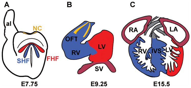
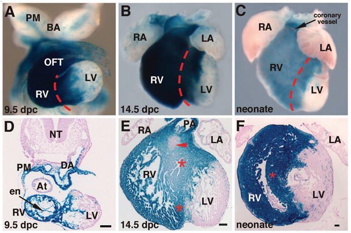
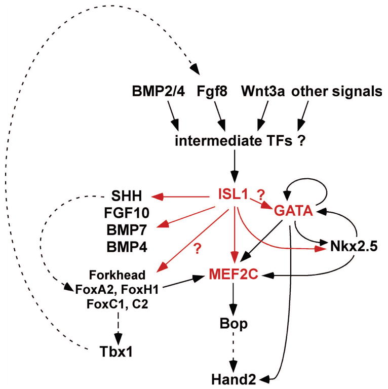
The heart is the first organ to form and function during vertebrate development and is absolutely essential for life. The left ventricle is derived from the classical primary or first heart field (FHF), while the right ventricle and outflow tract are derived from a distinct second heart field (SHF). The recent discovery of the SHF has raised several fundamental and important questions about how the two heart fields are integrated into a single organ and whether unique molecular programs control the development of the two heart fields. This review briefly highlights the contributions of the SHF to the developing and mature heart and then focuses primarily on our current understanding of the transcriptional pathways that function in the development of the SHF and its derivatives.


Figures

Fig. 1
Fig. 1
A schematic representation of three stages of heart development in the mouse. (A) A representation of a mouse embryo at embryonic day (E) 7.75 showing the location of three major populations of cells that contribute to the developing heart and outflow tract. The first heart field (FHF) is shown in red; the second heart field (SHF), which resides dorsal and medial to the FHF, is depicted in blue; and cardiac neural crest (NC) progenitors are shown at the dorsal neural tube in yellow. (B) A schematic of a mouse heart undergoing looping morphogenesis at E9 to E9.5. The FHF (red) contributes to the left ventricle. The SHF (blue) contributes primarily to the right ventricle (RV) and outflow tract (OFT). The sinus venosus (SV) or future atrial chamber is probably composed of FHF and SHF descendants. Two streams of plexinA2-positive cardiac neural crest cells entering the outflow tract are depicted in yellow. (C) A representation of a nearly mature embryonic mouse heart at E15.5. The free wall and majority of the left ventricle (LV) chamber is composed almost exclusively of cells derived from the FHF (red). The RV and interventricular septum (IVS) are composed primarily of cells descended from the SHF. The atria may be derived from a mixture of cells of FHF and SHF origins and the OFT is composed of cells of both NC and SHF origin. al, allantois; LA, left atrium; RA, right atrium.
Fig. 2
Fig. 2
Mef2c-AHF-Cre marks the SHF and its descendants. Transgenic mice harboring the mef2c-AHF-Cre transgene were crossed to ROSA26R lacZ reporter mice and progeny were collected at 9.5 days post coitus (dpc) (A, D), 14.5 dpc (B, E) or as neonates immediately after birth (C, F) and stained with X-gal as a measure of lacZ expression. Whole mount hearts are shown in (A–C). Transverse sections are shown in (D, F). A frontal section is shown in (E). These results support the idea that the right ventricle (RV), outflow tract (OFT), and interventricular septum (red asterisks and red dashed lines) are derived from the SHF. The red arrowhead in (E) marks the AV canal, which is unmarked by mef2c-AHF-Cre-expressing cells and their descendants. BA, branchial arches; At, atrium; DA, dorsal aorta; LA, left atrium; LV, left ventricle; NT, neural tube; PA, pulmonary artery; PM, pharyngeal mesoderm; RA, right atrium. Portions of this figure were derived with permission from Verzi et al., 2005 [21].
Fig. 3
Fig. 3
An ISL1-dependent transcriptional network for SHF development. In this model, the LIM-homeodomain transcription factor ISL1 functions as a key early regulator of SHF development by activating MEF2C and GATA transcription factors, which function as part of a core network for right ventricle and outflow tract development.

Similar articles

Cited by

References

    1. Brand T. Heart development: molecular insights into cardiac specification and early morphogenesis. Dev Biol. 2003;258:1–19. - PubMed
    1. Srivastava D. Making or breaking the heart: from lineage determination to morphogenesis. Cell. 2006;126:1037–48. - PubMed
    1. Olson EN. Gene regulatory networks in the evolution and development of the heart. Science. 2006;313:1922–7. - PMC - PubMed
    1. Garg V. Insights into the genetic basis of congenital heart disease. Cell Mol Life Sci. 2006;63:1141–8. - PMC - PubMed
    1. Srivastava D. Genetic assembly of the heart: implications for congenital heart disease. Annu Rev Physiol. 2001;63:451–69. - PubMed

Publication types

Substances

LinkOut - more resources