. 2007 Feb 6;17(3):193-202.
doi: 10.1016/j.cub.2006.12.040.

Intraflagellar transport protein 27 is a small G protein involved in cell-cycle control

Affiliations

Intraflagellar transport protein 27 is a small G protein involved in cell-cycle control

Hongmin Qin et al. Curr Biol. .

Abstract

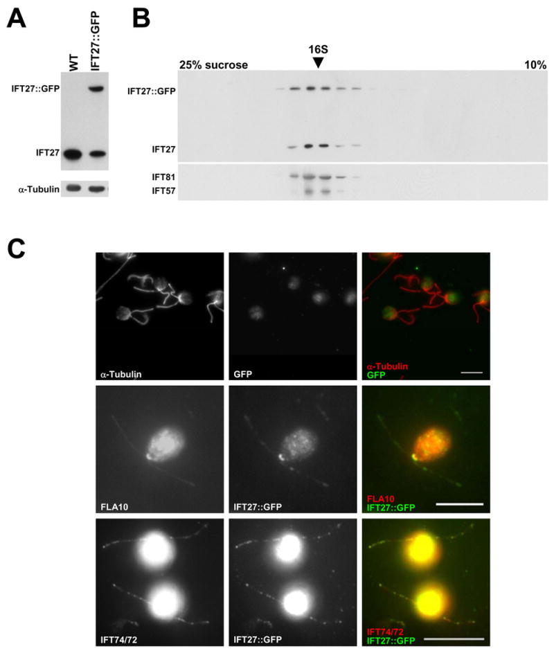
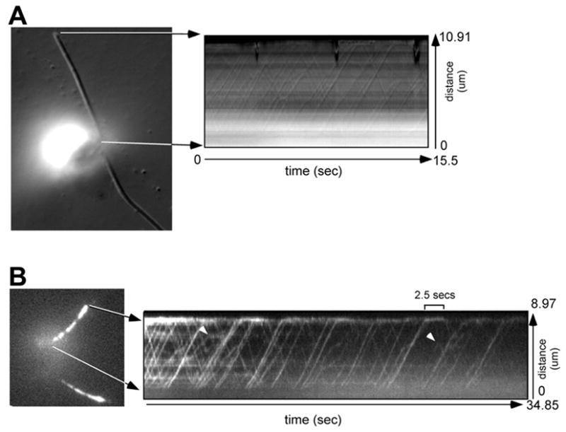
Background: Intraflagellar transport (IFT) is a motility process operating between the ciliary/flagellar (interchangeable terms) membrane and the microtubular axoneme of motile and sensory cilia. Multipolypeptide IFT particles, composed of complexes A and B, carry flagellar precursors to their assembly site at the flagellar tip (anterograde) powered by kinesin, and turnover products from the tip back to the cytoplasm (retrograde) driven by cytoplasmic dynein. IFT is essential for the assembly and maintenance of almost all eukaryotic cilia and flagella, and mutations affecting either the IFT motors or the IFT particle polypeptides result in the inability to assemble normal flagella or in defects in the sensory functions of cilia.

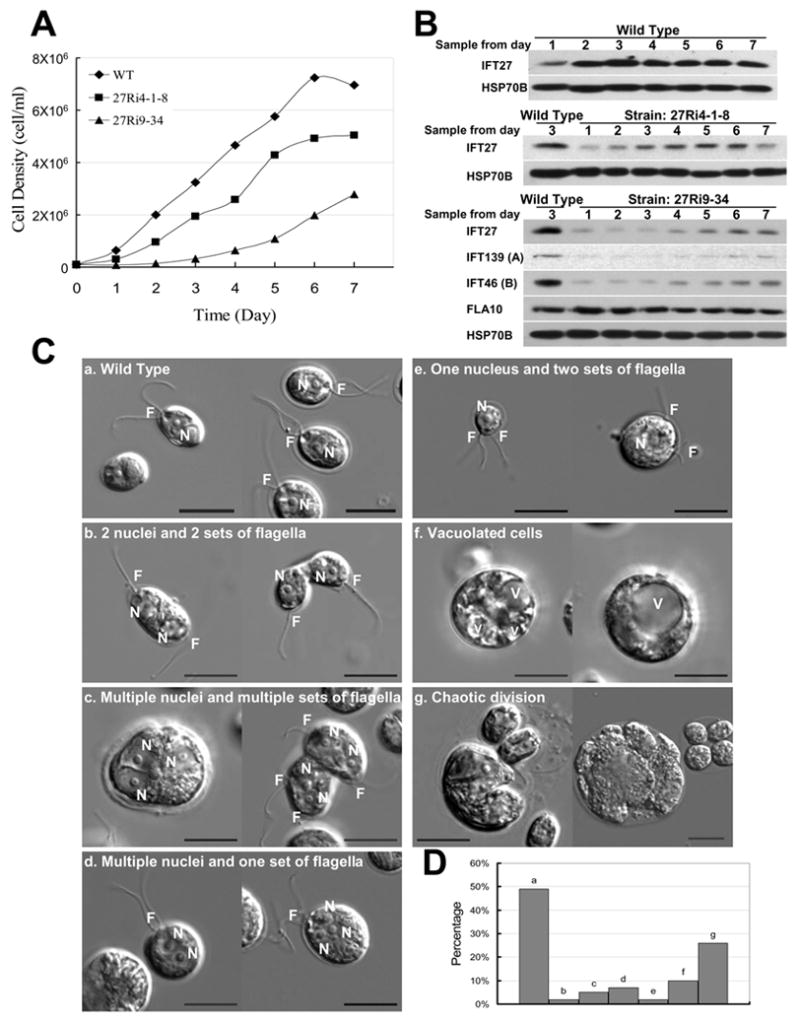
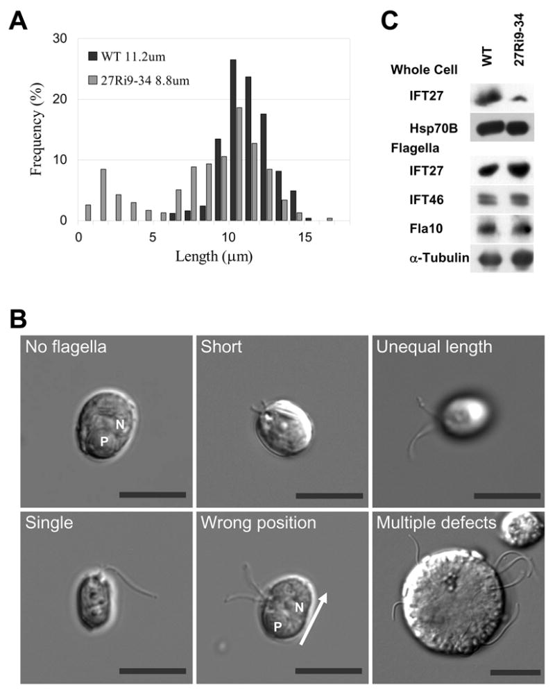
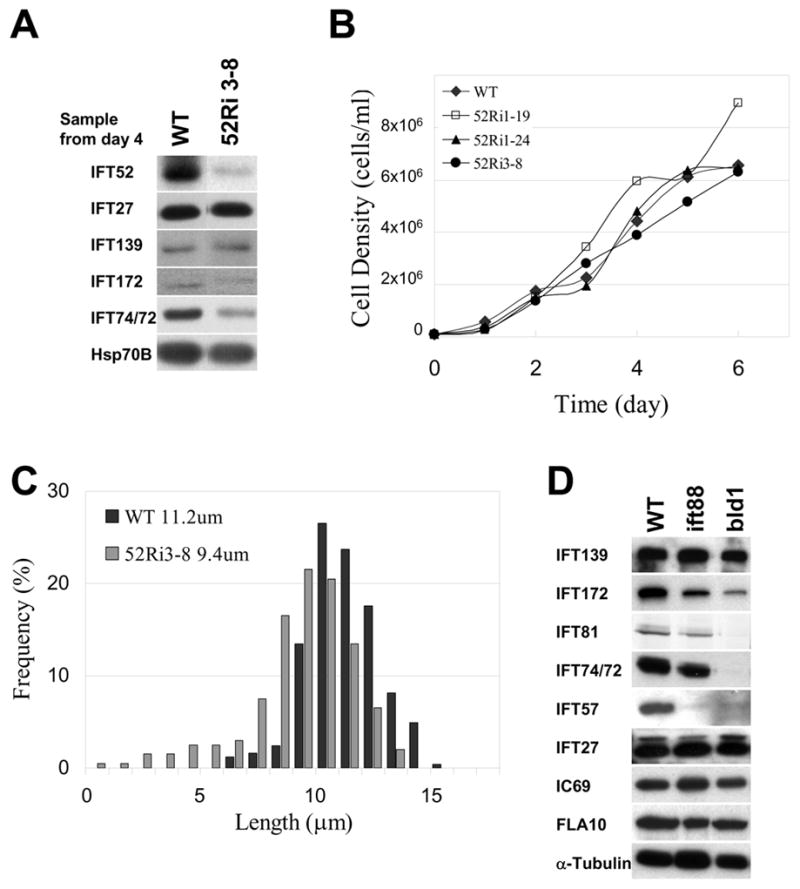
Results: We found that the IFT complex B polypeptide, IFT27, is a Rab-like small G protein. Reduction of the level of IFT27 by RNA interference reduces the levels of other complex A and B proteins, suggesting that this protein is instrumental in maintaining the stability of both IFT complexes. Furthermore, in addition to its role in flagellar assembly, IFT27 is unique among IFT polypeptides in that its partial knockdown results in defects in cytokinesis and elongation of the cell cycle and a more complete knockdown is lethal.

Conclusion: IFT27, a small G protein, is one of a growing number of flagellar proteins that are now known to have a role in cell-cycle control.


Figures

Figure 1
Figure 1. IFT27 is a Rab-like small G protein
IFT27 contains all five G domains required for GDP/GTP-binding and GTPase activities, which are conserved across the Ras superfamily of small G proteins. The IFT27 sequence is shown in darkened boxes and below the consensus sequences are listed where “X” is an unspecified amino acid and (X)n represents a sequence of indeterminate length. The possible lipid modification motif at the C-terminus is also highlighted.
Figure 2
Figure 2. IFT27::GFP can substitute for endogenous IFT27 and incorporate into IFT complexes in the flagella
(A) Immunoblots of whole flagella from wild type and IFT27::GFP transgenic cells probed for IFT27 showing two forms of IFT27 in the transgenic cells. Alpha-tubulin is included as a loading control. (B) Immunoblots of gradients of soluble flagellar proteins from IFT27::GFP transgenic cells probed with antibodies against several IFT polypeptides. IFT27::GFP sediments with the other IFT particle proteins, including endogenous IFT27. (C) Immunofluorescence of wild type and IFT27::GFP transgenic cells. Wild type cells show no flagellar or basal body labeling with the α-GFP antibody (top row). The second and third rows show that IFT27 co-localizes with FLA10 around the basal bodies and with IFT74/72 in the flagella. The IFT74/72 panel was brightened in PhotoShop to make the flagella readily visible. Scale Bars = 10um.
Figure 3
Figure 3. IFT27::GFP undergoes IFT
(A) The left panel is a DIC micrograph used for orientation. The kymograph (right panel) shows the trajectories of IFT particles moving bi-directionally in one flagellum of the cell. (B) The left panel is a fluorescence micrograph of a cell expressing IFT27::GFP. The kymograph (right panel) shows the movement of IFT27::GFP recorded by fluorescent microscopy. The lines in the kymographs were used to calculate the velocities of IFT particle or GFP movements. When IFT27::GFP reached the flagellar tip, it took up to a few seconds before reversing direction (one IFT27::GFP particle stayed at the tip for 2.5 secs). Some particles (arrow heads) reverse their direction before they reach the tip of the flagellum.
Figure 4
Figure 4. Knock down of IFT27 causes cell division defects
(A) Growth curves of wild type control (CC125) and two IFT27 RNAi knockdown clones (Ri 4-1-8 and Ri 9–34). (B) Immunoblots of whole cell extracts samples taken from the daily cultures in panel A. IFT27 was reduced in RNAi cells as were other IFT complex A and B proteins. FLA10 remained at the wild type level. HSP70B was used as a loading control. (C) DIC micrographs of wild type cells “a”, showing the normal cell shape and the position of nucleus “N” and flagella “F”; and IFT27 RNAi cells “b – g”, illustrating the major cell division defects seen in IFT27 knockdown cells. “V” = vacuole. (D) Plot of the percentages of the division defects for Ri 9–34 cells. The “a” bar represents the percentage of cells having one nucleus and one set of flagella. Many of the cells in this category had their flagella and nucleus positioned abnormally. “Set” refers to both pairs of flagella and unpaired singlet flagella. Scale bars = 10 μm.
Figure 5
Figure 5. Knockdown of IFT27 causes flagellar assembly and positioning defects
(A) Plots showing the length distribution of flagella from wild type and IFT27-depleted cells Ri 9–34 when the IFT27 level was 20% of wild type. Mean lengths are listed. (B) DIC micrographs illustrating the major flagellar defects in Ri 9–34. “P” = pyrenoid; “N” = nucleus; Bars = 10 μm. (C) Immunoblots comparing the levels of IFT proteins in whole cells and flagella of wild type and IFT27 knockdown cells. Although the level of IFT27 is greatly reduced in whole cells in the knockdown clone, the flagella of these cells have a normal complement of IFT proteins and FLA10.
Figure 6
Figure 6. Knock down of IFT52 causes flagellar assembly defects
(A) Immunoblots of wild type and IFT52 RNAi knockdown cells probed with antibodies against IFT polypeptides. Some IFT complex B proteins were reduced in the RNAi cells, but IFT27 and complex A protein IFT139 were not. HSP70B was included as a loading control. (B) Growth curves of wild type control (CC125) and three IFT52 RNAi knockdown clones. (C) Plots showing the length distribution of flagella in wild type and IFT52-depleted cells, 52Ri3–8, when the IFT52 level was 20% of wild type. Mean lengths are listed. (D) Immunoblots comparing the levels of IFT proteins in whole cells of wild type and mutants lacking IFT88 or IFT52 (bld1). Despite the reduction of most complex B proteins in these mutants, IFT27 and complex A proteins remained at wild type levels.

Similar articles

Cited by

References

    1. Dutcher SK. Elucidation of basal body and centriole functions in Chlamydomonas reinhardtii. Traffic. 2003;4:443–451. - PubMed
    1. Kozminski KG, Johnson KA, Forscher P, Rosenbaum JL. A motility in the eukaryotic flagellum unrelated to flagellar beating. Proc Natl Acad Sci USA. 1993;90:5519–5523. - PMC - PubMed
    1. Qin H, Diener DR, Geimer S, Cole DG, Rosenbaum JL. Intraflagellar transport (IFT) cargo: IFT transports flagellar precursors to the tip and turnover products to the cell body. J Cell Biol. 2004;164:255–266. - PMC - PubMed
    1. Cole DG, Diener DR, Himelblau AL, Beech PL, Fuster JC, Rosenbaum JL. Chlamydomonas kinesin-II-dependent intraflagellar transport (IFT): IFT particles contain proteins required for ciliary assembly in Caenorhabditis elegans sensory neurons. J Cell Biol. 1998;141:993–1008. - PMC - PubMed
    1. Scholey JM. Intraflagellar transport. Annu Rev Cell Dev Biol. 2003;19:423–443. - PubMed

Publication types

MeSH terms