. 2007 Feb 6;17(3):213-24.
doi: 10.1016/j.cub.2006.12.045.

A role for Cdc2- and PP2A-mediated regulation of Emi2 in the maintenance of CSF arrest

Affiliations

A role for Cdc2- and PP2A-mediated regulation of Emi2 in the maintenance of CSF arrest

Qiju Wu et al. Curr Biol. .

Abstract

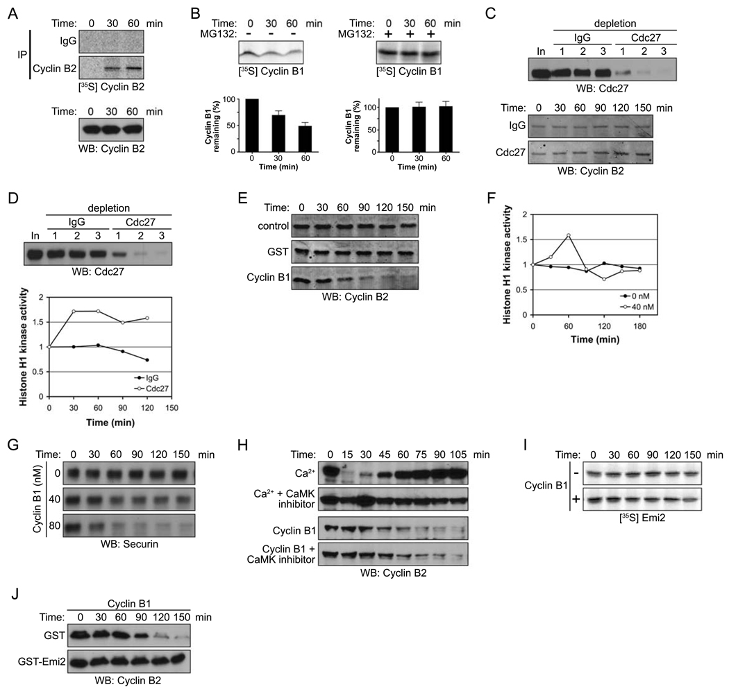
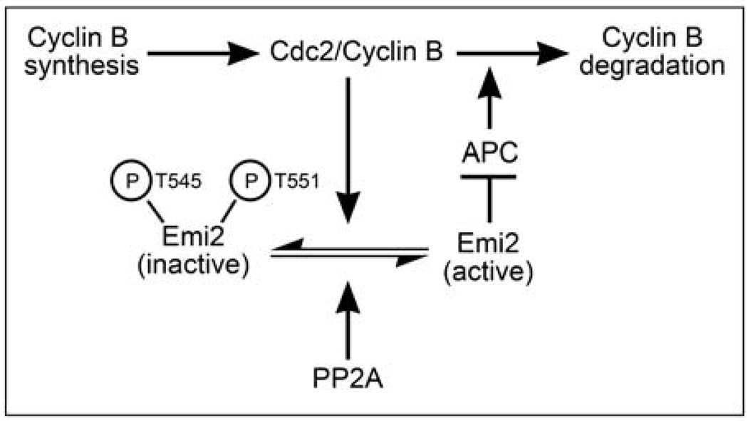
Background: Vertebrate oocytes are arrested in metaphase II of meiosis prior to fertilization by cytostatic factor (CSF). CSF enforces a cell-cycle arrest by inhibiting the anaphase-promoting complex (APC), an E3 ubiquitin ligase that targets Cyclin B for degradation. Although Cyclin B synthesis is ongoing during CSF arrest, constant Cyclin B levels are maintained. To achieve this, oocytes allow continuous slow Cyclin B degradation, without eliminating the bulk of Cyclin B, which would induce release from CSF arrest. However, the mechanism that controls this continuous degradation is not understood.

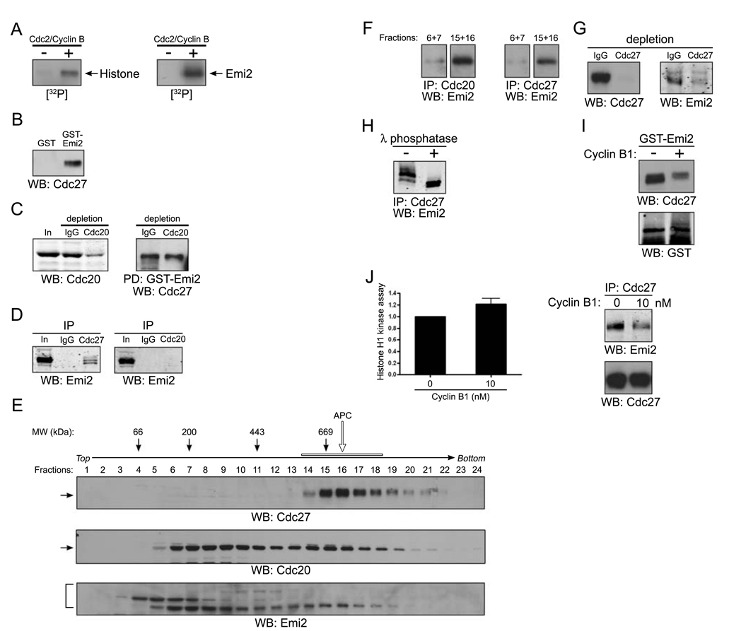
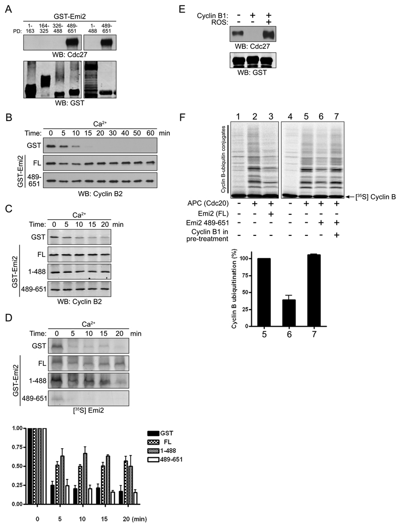
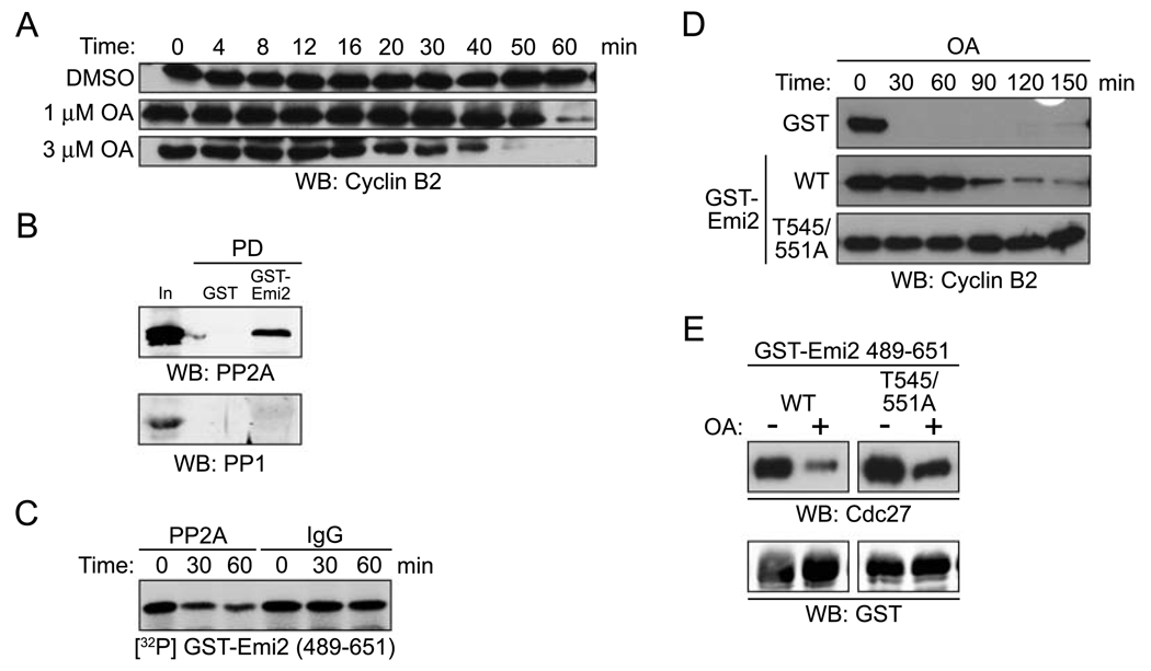
Results: We report here the molecular details of a negative feedback loop wherein Cyclin B promotes its own destruction through Cdc2/Cyclin B-mediated phosphorylation and inhibition of the APC inhibitor Emi2. Emi2 bound to the core APC, and this binding was disrupted by Cdc2/Cyclin B, without affecting Emi2 protein stability. Cdc2-mediated phosphorylation of Emi2 was antagonized by PP2A, which could bind to Emi2 and promote Emi2-APC interactions.

Conclusions: Constant Cyclin B levels are maintained during a CSF arrest through the regulation of Emi2 activity. A balance between Cdc2 and PP2A controls Emi2 phosphorylation, which in turn controls the ability of Emi2 to bind to and inhibit the APC. This balance allows proper maintenance of Cyclin B levels and Cdc2 kinase activity during CSF arrest.


Figures

Figure 1
Figure 1. Emi2 antagonizes Cdc2/Cyclin B-induced degradation of Cyclin B
(A) (Top): CSF-arrested Xenopus egg extracts were incubated in the presence of 35S-labeled methionine and cysteine. At the indicated times, endogenous Cyclin B2 protein was immunoprecipitated and newly synthesized Cyclin B2 was detected by autoradiography. (Bottom): Total Cyclin B2 levels were measured in a CSF extract at the indicated times by immunoblotting. (B) (Top): 35S-labeled Cyclin B1 was added into CSF extracts in the absence (−, left) or the presence (+, right) of MG132 (100 µM). Aliquots were withdrawn at the indicated times and the amount of Cyclin B1 was detected by autoradiography. (Bottom): The radiolabeled Cyclin B1 remaining at each time point was quantified. Error bars represent standard deviation of four measurements. (C) (Top): The APC was depleted from CSF extracts with three sequential incubations with IgG or Cdc27 antibodies. Samples from each extract were resolved by SDS-PAGE and immunoblotted with anti-Cdc27 antibodies. (Bottom): Samples were taken from control or APC (Cdc27) depleted extracts at the indicated times after incubation with energy regenerating mix at room temperature. The amount of Cyclin B2 was examined by immunoblotting (10 µg total protein was loaded per lane to clearly visualize the changes in endogenous Cyclin B levels). (D) (Top) The APC was depleted from CSF extracts as described in (C). (Bottom): After 30 min incubation at room temperature, the Cdc2/Cyclin B kinase activity in control or APC (Cdc27) depleted extracts was measured at the indicated times using Histone H1 kinase assays. The phosphorylation of Histone H1 was quantified, normalized and plotted. (E) Buffer (control), GST, or 40 nM Cyclin B1 was added to CSF extracts. At the indicated times, samples were withdrawn and endogenous Cyclin B2 levels were measured by immunoblotting. (F) At the indicated times, Cdc2/Cyclin B kinase activity in the CSF extracts was measured in the presence or absence of recombinant Cyclin B1 (40 nM) using Histone H1 as an exogenous substrate. The phosphorylation of Histone H1 was quantitated, normalized, and plotted. (G) Buffer (control) or Cyclin B1 (40 nM or 80 nM) was added to CSF extracts. At the indicated times, samples were withdrawn and Securin levels were measured by immunoblotting. (H) Ca2+ or recombinant Cyclin B1 was added into CSF extracts in the presence or absence of the CaMKII peptide inhibitor (281–309, 400 µM). Samples were taken at the indicated times and the amount of endogenous Cyclin B2 was measured by immunoblotting. (I) Radiolabeled Emi2 was added into CSF extracts supplemented with 40 nM recombinant Cyclin B1. The levels of Emi2 in the extract were examined at the indicated times by autoradiography. (J) Cyclin B1 was added into CSF extracts in the presence of GST or GST-Emi2. At the indicated times, samples were taken and the amount of endogenous Cyclin B2 was determined by immunoblotting.
Figure 2
Figure 2. Cdc2/Cyclin B disrupts the association of Emi2 and APC/Cdc20
(A) Histone H1 (left) or in vitro translated Emi2 (right) was mixed with [γ-32P] ATP in the presence or absence of recombinant Cdc2/Cyclin B kinase at room temperature for 10 min. The reactions were separated by SDS-PAGE and the phosphorylation of Histone H1 or Emi2 was monitored by autoradiography. (B) GST or GST-Emi2 proteins bound to glutathione Sepharose beads were incubated in CSF extracts. After incubation, the protein beads were pelleted, washed, and immunoblotted for the core APC component, Cdc27. (C) (Left): CSF extracts were depleted of Cdc20 by two consecutive incubations with Cdc20 antibody. The mock (IgG) and Cdc20 depleted extracts were resolved by SDS-PAGE and immunoblotted with anti-Cdc20 antibodies. (Right): GST-Emi2 linked to glutathione Sepharose was incubated in control or Cdc20 depleted extracts, retrieved, washed, and immunoblotted for Cdc27. (D) Control IgG, Cdc27 (left) or Cdc20 (right) antibodies were immobilized on Protein A Sepharose beads and incubated in CSF extracts. The beads were retrieved, washed and immunoblotted for Emi2. (E) CSF extracts was fractionated on a 5–30% sucrose gradient. Fractions were collected, analyzed by SDS-PAGE, and immunoblotted for Cdc27, Cdc20 and Emi2. The open arrow and bar indicate the APC complex. (F) Cdc20 (left) or Cdc27 (right) antibodies were immobilized on Protein A Sepharose beads and incubated in sucrose gradient fractions 6 and 7 (pooled) or 15 and 16 (pooled). After incubation, the beads were retrieved by centrifugation, washed, and immunoblotted for Emi2. (G) Cdc27 antibody was used to deplete CSF extract of the APC. IgG or Cdc27 depleted extracts were immunoblotted for Cdc27 (left) or Emi2 (right). (H) Emi2 antibodies immobilized on Protein A Sepharose beads were incubated in CSF extracts. After incubation, the beads were retrieved, washed and incubated in the presence (+) or the absence (−) of λ phosphatase before being resolved by SDS-PAGE and immunoblotting for Emi2. (I) GST-Emi2 linked to glutathione Sepharose was incubated in CSF extracts in the presence or absence of 80 nM recombinant Cyclin B1 proteins. After incubation, the protein beads were retrieved by centrifugation, washed, and immunoblotted for Cdc27 or GST. (J) (Left): Cdc2/Cyclin B kinase activity was measured in CSF extracts in the presence or absence of exogenous recombinant Cyclin B1 (10 nM) using Histone H1 as an exogenous substrate. The phosphorylation of Histone H1 was quantitated and plotted. Error bars represent standard deviation of three measurements. (Right): Cdc27 antibodies were immobilized on Protein A Sepharose beads and incubated in CSF extracts in the presence or absence of Cyclin B1 (10 nM). The beads were retrieved, washed and immunoblotted for Emi2 or Cdc27.
Figure 3
Figure 3. Cdc2/Cyclin B disrupts interactions between the C-terminal region of Emi2 and the APC
(A) GST-Emi2 protein fragments bound to glutathione Sepharose were incubated in CSF extracts, retrieved, washed, and immunoblotted for Cdc27 or GST. (B) GST or GST-Emi2 proteins (full-length (FL) or C-terminal (aa 489–651)) were added into CSF extracts supplemented with Ca2+. Aliquots removed at the indicated times were analyzed by SDS-PAGE and immunoblotted for Cyclin B2. (C) Emi2 full-length (FL), N-terminal (aa 1–488), or C-terminal (aa 489–651) GST proteins were added into CSF extracts supplemented with Ca2+. Aliquots were removed at the indicated times and immunoblotted for Cyclin B2. (D) (Top): 35S-labeled Emi2 was added to CSF extracts supplemented with GST or the indicated GST-Emi2 proteins. After Ca2+ addition, aliquots were removed at the indicated times and the amount of 35S-labeled Emi2 was examined by autoradiography. (Bottom): The amount of 35S-labeled Emi2 at each time point was quantified, normalized, and plotted. Error bars represent standard deviation of three replicates. (E) GST-Emi2 (aa 489–651) (4 µM) was incubated in CSF extracts in the presence or absence of 80 nM recombinant Cyclin B1 with or without the Cdc2 kinase inhibitor, roscovitine (ROS, 100 µM). GST-Emi2 was retrieved from the extracts by centrifugation, washed, and immunoblotted for Cdc27 or GST. (F) (Top): GST-Emi2 FL (lanes 1–3) or GST-Emi2 (aa 489–651, lanes 4–7) immobilized on glutathione Sepharose was incubated in CSF extracts in the presence or absence of excess recombinant Cyclin B1. The protein beads were retrieved and washed thoroughly and the proteins were then eluted using glutathione. Eluted proteins were then added to an in vitro APC assay and conversion of radiolabeled Cyclin B1 to ubiquitinated forms was monitored by SDS-PAGE and autoradiography. (Bottom): The amount of ubiquitin conjugated Cyclin B was quantified, normalized and plotted (lanes 5–7). Error bars represent standard deviation of four experiments.
Figure 4
Figure 4. Phosphorylation of Emi2 by Cdc2/Cyclin B at T545 and T551 disrupts APC binding and inhibition
(A) GST-Emi2 protein (aa 489–651) was incubated in CSF extracts in the absence (top) or presence (bottom) of recombinant Cyclin B1. After incubation, the protein was retrieved on glutathione Sepharose, resolved by SDS-PAGE, and the relevant bands were excised from the gel, trypsin digested, and analyzed by LC/MS mass spectrometry. The mass spectra of tryptic peptides were examined and the phosphorylated peptide (bottom: residues 545–556, [M2+H]2+ at m/z 649.6, retention time at 7.0 min) shows a 80 Da mass increase compared to the non-phosphorylated form (top: [M2+H]2+ at m/z 609.6, retention time at 7.8 min). Asterisks indicate tryptic peptides derived from GST protein. (B) The same experiment was repeated as in (A) with GST-Emi2 (aa 489–651) T545/551A protein in the presence of Cyclin B1. (Top): LC/MS extracted ion chromatograms (EIC) at m/z 1157.6 shows that unphosphorylated mutant peptide elutes at 6.8 min. (Middle): EIC at m/z 1237.6 shows that presumed monophosphorylated mutant peptide is not detected. (Bottom): EIC at m/z 1317.6 shows that presumed diphosphorylated mutant peptide is not detected. (C) GST, GST-Emi2 (aa 489–651) WT or GST-Emi2 (aa 489–651) T546/551A proteins were incubated in CSF extract supplemented with 80 nM recombinant Cyclin B1 protein in the presence of [γ-32P] ATP. Phosphorylation of the indicated proteins was examined by autoradiography at 0 and 10 min. (D) (Top): Cdc2 was depleted from CSF extracts by three consecutive incubations with recombinant His-p13 coupled to CnBr-activated Sepharose. Control and Cdc2-depleted extracts were immunoblotted with anti-Cdc2 antibody. (Bottom): GST-Emi2 (aa 489–651) WT protein was incubated in the depleted extracts supplemented with 80 nM recombinant Cyclin B1 in the presence of [γ-32P] ATP. The phosphorylation of GST-Emi2 (aa 489–651) WT protein was examined by autoradiography at 0 and 10 min. (E) GST, GST-Emi2 (aa 489–651) WT or GST-Emi2 (aa 489–651) T546/551A proteins were incubated with [γ-32P] ATP in the presence or absence of recombinant Cdc2/Cyclin B kinase for 10 min. The reactions were separated by SDS-PAGE and the phosphorylation of the indicated proteins was monitored by autoradiography. (F) GST-Emi2 protein (aa 489–651; WT, T545/551A, T545A, or T551A) linked to glutathione Sepharose was incubated in CSF extracts in the presence or absence of exogenous Cyclin B1. After incubation, the protein beads were retrieved by centrifugation, washed, and immunoblotted for Cdc27 or GST. (G) Recombinant Cyclin B1 was added into CSF extracts also supplemented with either GST, GST-Emi2 (aa 489–651) WT, or GST-Emi2 (aa 489–651) T545/551A proteins. At the indicated times, samples were withdrawn and immunoblotted for endogenous Cyclin B2. (H) MBP, MBP-Emi2 (aa 489–651) WT, or MBP-Emi2 (aa 489–651) T545/551A proteins were added into CSF extracts. Aliquots were removed at the indicated times and the Cdc2/Cyclin B kinase activity was measured by Histone H1 kinase assays. The phosphorylation of Histone H1 was measured, normalized, and plotted. (I) (Top): An in vitro APC assay was performed as described in Figure 3F using WT or T545/551A mutant Emi2 proteins (aa 489–651). (Bottom): The amount of ubiquitin conjugated Cyclin B was quantified, normalized, and plotted (lanes 2–6). Error bars represent standard deviation of four experiments.
Figure 5
Figure 5. An okadaic acid-sensitive phosphatase keeps Emi2 active to maintain CSF arrest
(A) GST-Emi2 (FL) protein beads were incubated in CSF extracts to bind Cdc27. The beads were retrieved, washed, and then incubated in fresh buffer containing MgCl2 and ATP in the presence of DMSO (−), okadaic acid (OA), recombinant Cdc2/Cyclin B1 or in combination as indicated. After incubation, the protein beads were pelleted, washed, and immunoblotted for Cdc27 and GST. (B) Histone H1 protein was incubated with [γ-32P] ATP and recombinant Cdc2/Cyclin B kinase in the presence or absence of or OA for 10 min. The reactions were separated by SDS-PAGE and the phosphorylation of Histone H1 was monitored by autoradiography. (C) DMSO or 5 µM OA was added to CSF extracts. Samples were taken at the indicated times and the endogenous Cyclin B2 levels were examined by immunoblotting. (D) CSF extracts were incubated with or without OA and the Cdc2 kinase activity of the extracts was examined using Histone H1 as a substrate. The reactions were separated by SDS-PAGE and the phosphorylation of Histone H1 was monitored by autoradiography. (E) GST-Emi2 (FL) protein linked to glutathione Sepharose was incubated in CSF extracts supplemented with either DMSO or OA. After incubation, the protein beads were retrieved, thoroughly washed, and transferred into fresh CSF extracts supplemented with OA. Samples were taken at the indicated times to monitor Cyclin B2 levels by immunoblotting. (F) 35S-labeled Cyclin B1 and Emi2 proteins were synthesized in vitro and added to CSF extracts. After 15 min incubation, Ca2+, DMSO or OA (or both Ca2+ and OA) were added. Aliquots were removed at the indicated times and the degradation or gel mobility shifting of both Cyclin B1 and Emi2 were examined by autoradiography using a phosphorimager.
Figure 6
Figure 6. Emi2 associates with PP2A acting in opposition of Cdc2/Cyclin B kinase activity
(A) DMSO, 1 or 3 µM OA was added to CSF extracts. Aliquots were removed at the indicated times and immunoblotted for endogenous Cyclin B2. (B) GST or GST-Emi2 (FL) protein beads were incubated in CSF extracts. After incubation, the protein beads were retrieved, washed, and immunoblotted for PP2A (top) and PP1 (bottom). (C) IgG or PP2A antibody was immobilized on Protein A Sepharose beads and incubated in CSF extracts. The beads were retrieved, washed and mixed with GST-Emi2 (aa 489–651) WT protein that was pre-phosphorylated with recombinant Cdc2/Cyclin B kinase in the presence of [γ-32P] ATP. The phosphorylation of GST-Emi2 (aa 489–651) WT was examined by autoradiography after 0, 30 and 60 min. (D) OA was added into CSF extracts supplemented with either GST, GST-Emi2 WT, or GST-Emi2 T545/551A proteins. At the indicated times, samples were withdrawn and immunoblotted for endogenous Cyclin B2. (E) GST-Emi2 protein beads (aa 489–651; WT or T545/551A) were incubated in CSF extracts in the presence of DMSO (−) or OA. After incubation, the protein beads were retrieved, washed, and immunoblotted for Cdc27 and GST.
Figure 7
Figure 7. Cyclin B levels are regulated by Cdc2 and PP2A modulation Emi2 during a CSF arrest
Cyclin B proteins are continually synthesized and degraded during a CSF arrest. When Cyclin B levels become elevated due to ongoing synthesis, the resulting increase in Cdc2 kinase activity tilts the balance between Cdc2/Cyclin B and PP2A, allowing for the phosphorylation of Emi2. This promotes the dissociation of Emi2 from the APC, thus activating a proportion of APC, and allowing for some Cyclin B degradation. Degradation of Cyclin B lowers Cdc2 kinase activity, allowing PP2A to predominate, thus shifting the balance towards dephosphorylated Emi2, which can inhibit the APC to block further Cyclin B degradation.

References

    1. Masui Y, Markert CL. Cytoplasmic control of nuclear behavior during meiotic maturation of frog oocytes. J Exp Zool. 1971;177:129–145. - PubMed
    1. Sagata N, Watanabe N, Vande Woude GF, Ikawa Y. The c-mos proto-oncogene product is a cytostatic factor responsible for meiotic arrest in vertebrate eggs. Nature. 1989;342:512–518. - PubMed
    1. Haccard O, Sarcevic B, Lewellyn A, Hartley R, Roy L, Izumi T, Erikson E, Maller JL. Induction of metaphase arrest in cleaving Xenopus embryos by MAP kinase. Science. 1993;262:1262–1265. - PubMed
    1. Bhatt RR, Ferrell JE., Jr The protein kinase p90 rsk as an essential mediator of cytostatic factor activity. Science. 1999;286:1362–1365. - PubMed
    1. Gross SD, Schwab MS, Lewellyn AL, Maller JL. Induction of metaphase arrest in cleaving Xenopus embryos by the protein kinase p90Rsk. Science. 1999;286:1365–1367. - PubMed

Publication types

MeSH terms

LinkOut - more resources