

Localized tufts of fibrils on Staphylococcus epidermidis NCTC 11047 are comprised of the accumulation-associated protein
- PMID: 17277069
- PMCID: PMC1855787
- DOI: 10.1128/JB.00952-06
Localized tufts of fibrils on Staphylococcus epidermidis NCTC 11047 are comprised of the accumulation-associated protein
Abstract
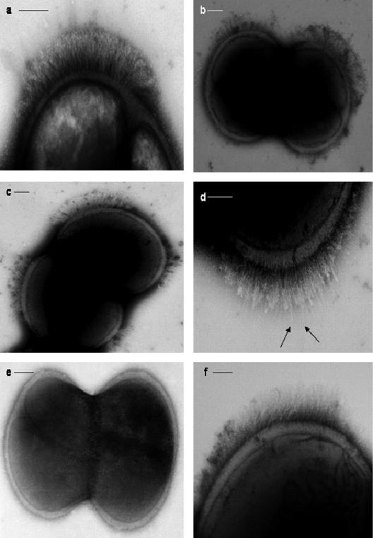
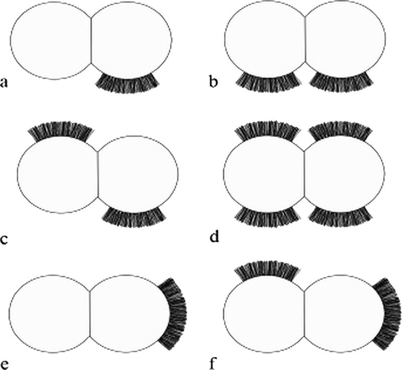
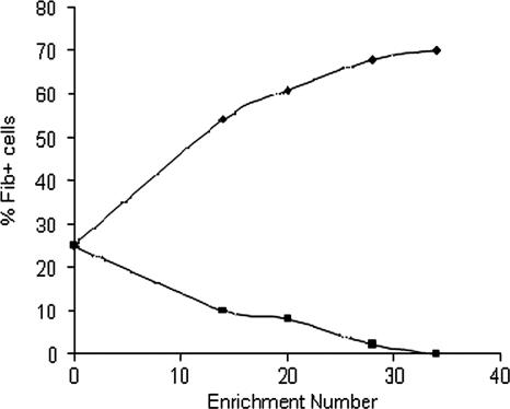
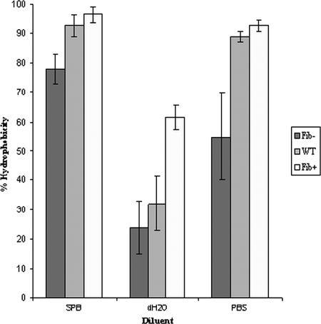
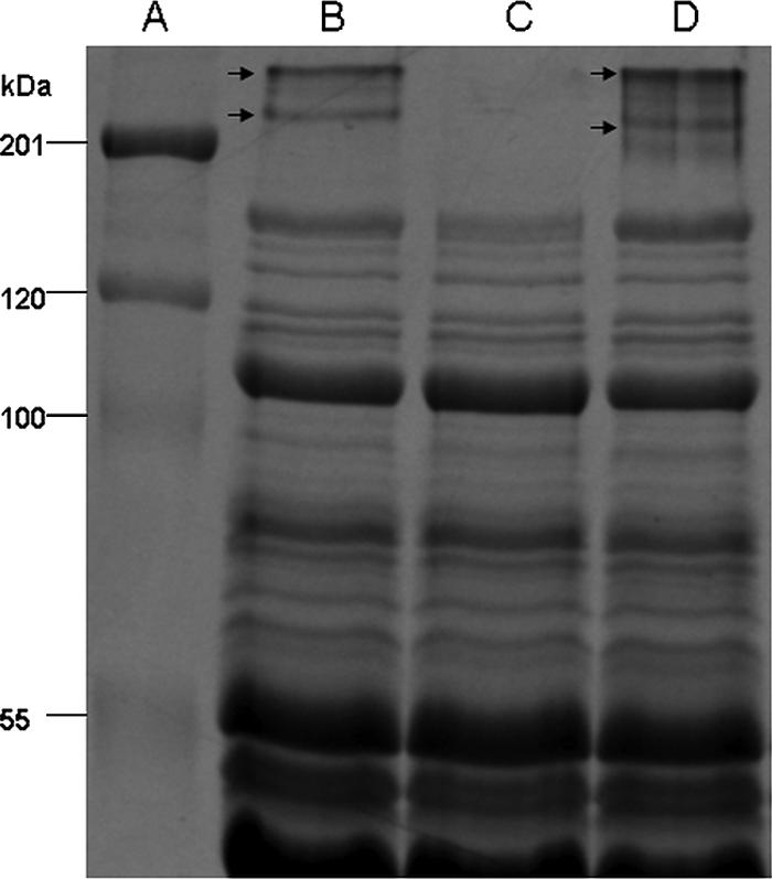
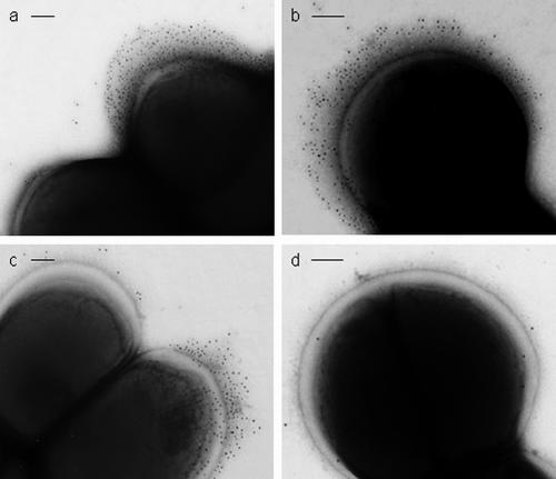
Staphylococcus epidermidis is both a human skin commensal and an opportunistic pathogen, causing infections linked to implanted medical devices. This paper describes localized tufts of fibrillar appendages on a subpopulation (25%) of wild-type (WT) S. epidermidis NCTC 11047 cells. The fibrils (122.2 +/- 10.8 nm long) are usually in a lateral position on the cells. Fibrillar (Fib(+)) and nonfibrillar (Fib(-)) subpopulations were separated (enriched) by 34 sequential partitions of WT cells between a buffer phase and a hexadecane phase. Following enrichment, hydrophobic cells from the hexadecane phase comprised 70% Fib(+) cells and the less hydrophobic cells from the buffer phase entirely comprised Fib(-) cells. The Fib(+) and Fib(-) subpopulations did not revert on subculture (34 times) on solid medium. Sodium dodecyl sulfate-polyacrylamide gel electrophoresis of cell surface proteins from WT, Fib(+), and Fib(-) cells revealed two high-molecular-mass proteins (280 kDa and 230 kDa) on the WT and Fib(+) cells that were absent from the Fib(-) cells. Amino acid sequencing revealed that fragments of both the 280- and 230-kDa proteins had 100% identity to the accumulation-associated protein (Aap). Aap is known to cause biofilm formation if it is truncated by loss of the terminal A domain. Immunogold staining with anti-Aap antibodies labeled tuft fibrils of the WT and Fib(+) cells but not the cell surface of Fib(-) cells. The tufts were labeled with N-terminally directed antibodies (anti-A domain), showing that the fibrillar Aap was not truncated on the cell surface. Thus, the presence of full-length Aap correlated with the low biofilm-forming abilities of both WT and Fib(+) S. epidermidis NCTC 11047 populations. Reverse transcription-PCR showed that aap was transcribed in both Fib(+) and Fib(-) cells. We therefore propose that full-length Aap is expressed on cells of S. epidermidis NCTC 11047 as tufts of short fibrils and that fibril expression is regulated at a posttranscriptional level.
Figures
References
-
- Arciola, C. R., S. Gamberoni, D. Capoccia, L. Visai, P. Speziale, L. Baldassarri, and L. Montanaro. 2005. A multiplex PCR method for the detection of all five individual genes of ica locus in Staphylococcus epidermidis. A survey on 400 clinical isolates from prosthesis-associated infections. J. Biomed. Mater. Res. A 75:408-413. - PubMed
-
- Bowden, M. G., W. Chen, J. Singvall, Y. Xu, S. J. Peacock, V. Valtulina, P. Speziale, and M. Hook. 2005. Identification and preliminary characterization of cell-wall-anchored proteins of Staphylococcus epidermidis. Microbiology 151:1453-1464. - PubMed
-
- Busscher, H. J., P. S. Handley, P. G. Rouxhet, L. M. Hesketh, and H. C. van der Mei. 1991. The relationship between structural and physicochemical surface properties of tufted Streptococcus sanguis strains, p. 317-338. In P. G. Rouxhet (ed.), Microbial cell surface analysis, structural and physicochemical methods. VCH Publishers Inc., New York, NY.
-
- Christensen, G. D., A. L. Bisno, J. T. Parisi, B. McLaughlin, M. G. Hester, and R. W. Luther. 1982. Nosocomial septicaemia due to multiply antibiotic-resistant Staphylococcus epidermidis. Ann. Intern. Med. 96:1-10. - PubMed
Publication types
MeSH terms
Substances
LinkOut - more resources
Full Text Sources
Other Literature Sources
Molecular Biology Databases
Miscellaneous
