. 2007 Feb 13;104(7):2181-6.
doi: 10.1073/pnas.0607052104. Epub 2007 Feb 2.

Direct measurement of force generation by actin filament polymerization using an optical trap

Affiliations

Direct measurement of force generation by actin filament polymerization using an optical trap

Matthew J Footer et al. Proc Natl Acad Sci U S A. .

Abstract

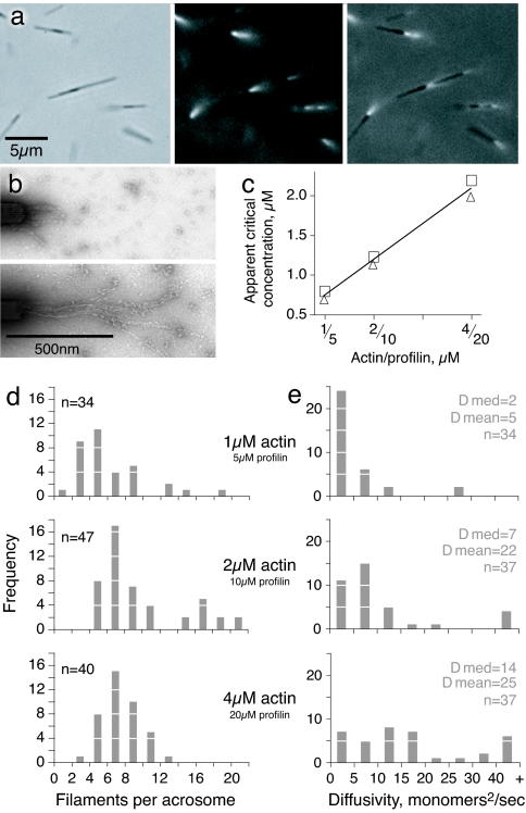
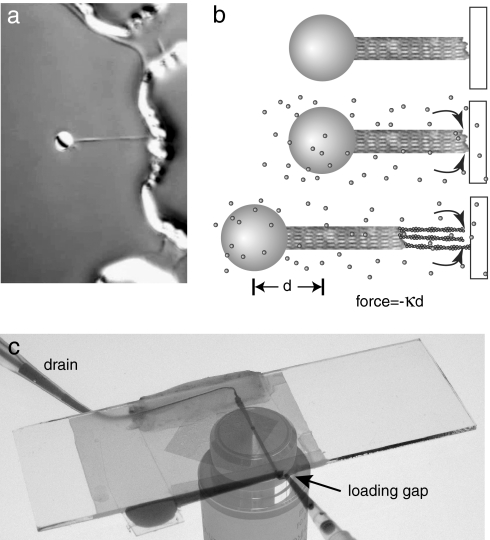
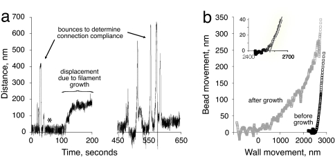
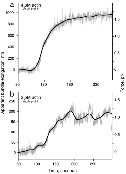
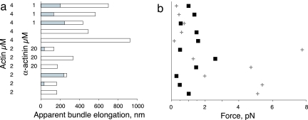
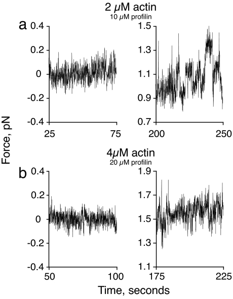
Actin filament polymerization generates force for protrusion of the leading edge in motile cells. In protrusive structures, multiple actin filaments are arranged in cross-linked webs (as in lamellipodia or pseudopodia) or parallel bundles (as in filopodia). We have used an optical trap to directly measure the forces generated by elongation of a few parallel-growing actin filaments brought into apposition with a rigid barrier, mimicking the geometry of filopodial protrusion. We find that the growth of approximately eight actin parallel-growing filaments can be stalled by relatively small applied load forces on the order of 1 pN, consistent with the theoretical load required to stall the elongation of a single filament under our conditions. Indeed, large length fluctuations during the stall phase indicate that only the longest actin filament in the bundle is in contact with the barrier at any given time. These results suggest that force generation by small actin bundles is limited by a dynamic instability of single actin filaments, and therefore living cells must use actin-associated factors to suppress this instability to generate substantial forces by elongation of parallel bundles of actin filaments.


Conflict of interest statement

The authors declare no conflict of interest.

Figures

Fig. 1.
Fig. 1.
Actin filament growth from isolated Limulus acrosomal bundles. (a) Limulus acrosomal bundle fragments (≈5 μm long) were incubated with rhodamine-actin. Small tufts of fluorescent actin filaments are seen growing from the barbed end in this sequence of phase-contrast, rhodamine, and overlay images. Notice that some acrosomes form clusters. (b) Electron micrographs of negatively stained filaments grown from acrosomal bundles with 4 μM monomeric actin and 20 μM profilin. (Upper) Ten seconds of growth. (Lower) Thirty seconds of growth. (c) Apparent actin critical concentration measured for different concentrations of profilin (actin/profilin ratio 1:5 for all experiments). Duplicate points show results of independent experiments. (d) The number of filaments per acrosome, counted during the first 40 s of growth. The difference in the number of filaments per acrosome at different time points was not significant. (e) Diffusivity in length of the actin filaments in the three assay conditions over the first 40 s of growth. The complete set of diffusivity data are presented in SI Fig. 8.
Fig. 2.
Fig. 2.
Experimental setup. (a) Bead with attached Limulus acrosomal bundle held in keyhole trap and brought next to microfabricated wall structure. Bead is 2 μm in diameter. (b) Schematic showing the sequence of events for an experiment. (Top) The bead-acrosome construct is positioned a few nanometers away from a barrier. (Middle) Actin monomers are introduced into the flow cell. (Bottom) Filaments grow from the barbed end of the acrosomal bundle and force the bead away from the wall. The force is directly proportional to the distance d. (c) Flow cell used in the experiments. The slide is on top and the coverslip (with diamond-shaped patterned area) is at the bottom. Dye shows the fluid path from reservoir to drain. The microscope objective was photographed separately and added for clarity.
Fig. 3.
Fig. 3.
Displacement of bead in laser trap caused by actin filament growth. (a) Positional data, raw trace. The zero-to-200-s portion shows initial “bounces” to characterize mechanical behavior of the bead and acrosomal bundle construct. Asterisk indicates time when actin was added. After growth reached a plateau, the bead and bundle were pulled away from the wall, and then the series of bounces were repeated. This particular experimental trace was not used for force measurements. (b) Bounce data from a represented as bead displacement as a function of deliberate movement of the wall, before and after actin filament elongation. The zero point was set to coincide with the first contact of the elongated filaments. Using this zero point the stage would have had to move ≈2,530 nm to make contact before the addition of actin.
Fig. 4.
Fig. 4.
Force measurement for actin filament growth with monomeric actin at 2 (b) and 4 (a) μM. Gray trace shows raw data, and the black line is the best-fit curve using the locally weighted least-squares method (46).
Fig. 5.
Fig. 5.
Filament lengths and forces necessary to buckle the elongating filaments. (a) Summary of final filament length determined by summing the distance the bundle tip was held away from the wall (shaded regions) and the filament elongation measured from the bead displacement (unshaded region) for 11 experiments. (b) Observed average force at stall (■) determined from bead displacement, compared with the force necessary to buckle a single filament (cross) as determined from the length in a. Vertical axis is as in a.
Fig. 6.
Fig. 6.
Noise characteristics of experimental traces before growth (Left) and during the plateau (Right) in the presence of 2 (a) and 4 (b) μM actin.

References

    1. Inoue S, Salmon ED. Mol Biol Cell. 1995;6:1619–1640. - PMC - PubMed
    1. Theriot JA. Traffic. 2000;1:19–28. - PubMed
    1. Mogilner A. Curr Opin Cell Biol. 2006;18:32–39. - PubMed
    1. Dogterom M, Kerssemakers JWJ, Romet-Lemonne G, Janson ME. Curr Opin Cell Biol. 2005;17:67–74. - PubMed
    1. Hill TL, Kirschner MW. Int Rev Cytol. 1982;78:1–125. - PubMed

Publication types