. 2007 Jun;28(6):1163-70.
doi: 10.1093/carcin/bgm025. Epub 2007 Feb 2.

Nucleophosmin suppresses oncogene-induced apoptosis and senescence and enhances oncogenic cooperation in cells with genomic instability

Affiliations

Nucleophosmin suppresses oncogene-induced apoptosis and senescence and enhances oncogenic cooperation in cells with genomic instability

June Li et al. Carcinogenesis. 2007 Jun.

Abstract

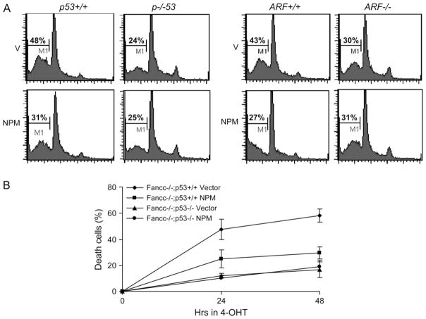
Cells from patients with genomic instability syndromes have high predisposition to cancer. However, little is known about whether these mutant cells have high susceptibility to oncogenic transformation. We have tested the hypothesis that a defect in maintaining genome integrity is necessary but not sufficient alone for oncogenic transformation and needs to collaborate with other signals in order to produce full oncogenic transformation. Using genetically matched primary cells deficient for the Fanconi complementation group C gene (Fancc) and the ataxia telangiectasia mutated gene (Atm), we found that certain forms of oncogenic activation and cooperation require a combination of genomic instability with increased expression of nucleophosmin (NPM) to prevent oncogenic stress-induced apoptosis or senescence. Intriguingly, co-expression of c-Myc and NPM leads to a synergistic increase in the proliferation rate in Fancc-/- or Atm-/- cells. Analysis of p53 stabilization and activation by c-Myc demonstrates that over-expression of NPM significantly reduces the accumulation of the activated p53 but not the stability of p53. Moreover, NPM is shown to enhance transforming activity of co-expressed Myc and Ras in wild-type and, to a greater degree, in Fancc-/- or Atm-/- cells, suggesting a role in oncogenic cooperation. Finally, a partial knockdown of NPM is sufficient to cause massive apoptosis in Fancc-/- or Atm-/- cells co-expressing c-Myc and Ras while sparing untransformed cells. Our study demonstrates a novel mechanism of NPM tumorigenesis by establishing NPM as a crucial inhibitor of oncogene-induced apoptosis and senescence.


Conflict of interest statement

Conflict of Interest Statement: None declared.

Figures

Fig. 1
Fig. 1
Over-expression of NPM suppresses c-Myc-induced senescence and apoptosis in Fancc−/− and Atm−/− cells. (A) Expression of retrovirally encoding proteins in Fancc−/− and Atm−/− cells. WT, Fancc−/− or Atm−/− MEFs were infected at passage 2 with pMSCVpuro empty vector (V) or pMSCVpuro-NPM retrovirus. After 72 h puromycin selection, the cells were split and infected with the control vector (control) or MycER viruses. Forty-eight hours after transduction, cells were cultured in the presence of 125 nM 4-hydroxy tamoxifen. Western blot analysis shows expression of the expected Flag-tagged NPM, endogenous NPM and c-Myc proteins. Fifty micrograms of total proteins were loaded in each lane, and equal loading was ensured by probing the blot with anti-β-actin. (B) NPM inhibits c-Myc-induced senescence. Cells described in (A) were further cultured for 48 h and stained for SA-β-gal. The graphs on the right are percentages of the cells stained positive for SA-β-gal quantified by counting a total of 100 cells in random fields on a slide. The data represent the mean ± SD of three independent experiments. (C) Cells described in (A) were analyzed for subdiploid DNA content by flow cytometry 24 h after transfer to medium containing 125 nM 4-hydroxy tamoxifen. Data shown represent mean ± SD of three independent experiments. *Statistical significance between Myc + V and Myc + NPM samples at P < 0.05. (D) Effect of NPM over-expression on proliferation of Myc-activated WT, Fancc−/− or Atm−/− cells. Cells synchronized at G0 were cultured for 24 h in normal growth medium supplemented with 10 μM BrdU, and level of BrdU incorporation was determined by flow cytometry. Data represent mean ± SD of three experiments. *Statistical significance between Myc + V and Myc + NPM samples at P < 0.05.
Fig. 2
Fig. 2
Over-expression of NPM inhibits Myc-induced p53 activation. Genetically matched WT and Fancc−/− (A) or WT and Atm−/− (B) MEFs were infected at passage 2 with pMSCVpuro empty vector (V) or pMSCVpuro-NPM retrovirus. After 72 h puromycin selection, the cells were split and infected with the control vector (control) or MycER viruses. Forty-eight hours after transduction, cells were cultured in the presence of 125 nM 4-hydroxy tamoxifen (4-OHT). Whole cell extracts were then prepared and 50 μg of total proteins from each sample was analyzed by western blot for expression of p53Ser15, p53, p21WAF1, NPM and c-Myc. β-Actin was used as a loading control. (C) WT and Fancc−/− cells were infected at passage 2 with pMSCVpuro empty vector or pMSCVpuro-NPM retrovirus. After 72 h puromycin selection, the cells were infected with MycER viruses. Forty-eight hours after transduction, cells were cultured in the presence or absence of 125 nM 4-hydroxy tamoxifen for 48 h. Whole cell extracts were then prepared and 100 μg of total proteins from each sample was analyzed by western blot for expression of γH2AX and p53Ser15. β-Actin was used as a loading control. (D) Cells described in (C) were treated with mitomycin C (0.5 μM) for 24 h in the presence of 125 nM 4-hydroxy tamoxifen for 48 h. Whole cell extracts were then prepared and 100 μg of total proteins from each sample was analyzed by western blot for expression of γH2AX and p53Ser15. β-Actin was used as a loading control.
Fig. 3
Fig. 3
NPM inhibits Myc-induced apoptosis requires a functional p53–ARF pathway. (A) Effect of NPM over-expression on Myc-induced apoptosis in p53- and ARF-null cells. Genetically matched p53 or ARF WT and null MEFs were infected with pMSCVpuro empty vector (V) or pMSCVpuro-NPM retrovirus. After 72 h puromycin selection, the cells were infected with the MycER viruses. Forty-eight hours after transduction, cells were cultured in the presence of 125 nM 4-hydroxy tamoxifen (4-OHT) for 24 h and analyzed for subdiploid DNA content by flow cytometry. (B) p53-dependent cell death in Fancc−/− cells expressing activated c-Myc. p53+/+, Fancc−/− or p53−/− and Fancc−/− MEFs were infected with pMSCVpuro empty vector (V) or pMSCVpuro-NPM retrovirus. After 72 h puromycin selection, the cells were infected with the MycER viruses. Forty-eight hours after transduction, cells were cultured in the presence of 125 nM 4-hydroxy tamoxifen and cell viability was measured at 0, 24 and 48 h after transfer to 4-hydroxy tamoxifen-containing medium.
Fig. 4
Fig. 4
c-Myc-induced NPM–ARF and NPM–p53 interactions in Fancc−/− and Atm−/− cells. WT or Fancc−/− in p53 WT or null background (A) or Atm WT or null (B) MEFs were infected with pMSCVpuro-NPM retroviruses. After 72 h puromycin selection, the cells were infected with the control vector (control) or MycER viruses. Forty-eight hours after transduction, cells were cultured in the presence of 125 nM 4-hydroxy tamoxifen for 24 h. Whole cell extracts (500 μg in 500 μl) were incubated with 20 μl of the anti-FLAG (M2) antibodies conjugated to agarose beads. The input (left panel; 50 μg per lane) and anti-Flag immunocomplexes were analyzed by immunoblotting with antibodies specific for p53, p19ARF or Flag peptides. (C) WT or Fancc−/− cells were infected with pMSCVpuro-NPM retroviruses. After 72 h puromycin selection, the cells were infected with the control vector (control) or MycER viruses. Forty-eight hours after transduction, cells were cultured in the presence of 125 nM 4-hydroxy tamoxifen for 24 h. Whole cell extracts (500 μg in 500 μl) were incubated with 20 μl of the anti-p53 (DO-1) antibodies conjugated to agarose beads. The input (left panel; 50 μg per lane) and anti-p53 immunocomplexes were analyzed by immunoblotting with antibodies specific for NPM or p53. Note that one-fifth of total immunoprecipitates from c-Myc-expressing cells were loaded. IgG, IgG-heavy chain.
Fig. 5
Fig. 5
NPM over-expression enhances oncogenic cooperation in Fancc−/− and Atm−/− primary cells. (A) WT or Fancc−/− MEFs were infected at passage 2 with pMSCVpuro-NPM retroviruses. After 72 h puromycin selection, the cells were split and infected with the control vector (control) or MycER, H-ras-G12Vor both MycER and H-ras-G12V viruses. Forty-eight hours after transduction, 1 × 10⁴ cells were plated in each well of six-well plate in triplicate in semi-solid medium containing 125 nM 4-hydroxy tamoxifen. Two weeks after plating, colonies were counted and photographed. The numbers at upper right corners are colonies per plate. (B) WT or Atm−/− MEFs were infected at passage 2 with pMSCVpuro-NPM retroviruses. After 72 h puromycin selection, the cells were split and infected with the control vector (control), MycER, H-ras-G12V or both MycER and H-ras-G12V viruses. Forty-eight hours after transduction, 1 × 10⁴ cells were plated in each well of six-well plate in triplicate in semi-solid medium containing 125 nM 4-hydroxy tamoxifen. Two weeks after plating, colonies were counted and data represent mean ± SD of three experiments. *Statistical significance between Myc + Ras and NPM + Myc + Ras samples at P < 0.05. (C) Foci formation. Cells (1 × 10⁴) described in (A) and (B) were plated and cultured in growth medium containing 125 nM 4-hydroxy tamoxifen for two weeks. Then the cells were fixed, stained and colonies were counted. Data represents mean ± SD of three experiments performed in triplicate. *Statistical significance between Myc + Ras and NPM + Myc + Ras samples at P < 0.05.
Fig. 6
Fig. 6
Partial NPM inactivation by RNA interference sensitizes Fancc−/− and Atm−/− cells to oncogene-induced apoptosis. (A) Levels of NPM protein in genetically matched Fancc or Atm MEFs were infected at passage 2 with pMSCVpuro-NPM retroviruses. After 72 h puromycin selection, the cells were co-infected with the MycER and H-ras-G12V viruses. Forty-eight hours after transduction, cells were transfected with NPM siRNA (50, 100 and 150 nM) or the scramble siRNA (150 nM). Another 36 h later, the cells were analyzed by western blotting for NPM expression level, which was also normalized for loading by probing the western blot with antibody for β-actin. (B) Partial NPM silencing causes transformed Fancc−/− and Atm−/− cell death as analyzed by measurement of fractional subdiploid DNA content after staining with propidium iodide and analysis by flow cytometry. Cells described in (A) were cultured in the presence of 125 nM 4-hydroxy tamoxifen for additional 24 h. Data reflect means ± SD of two independent experiments performed. *Statistical significance between Scramble and siNPM samples at P < 0.05.

References

    1. Strasser A, et al. Novel primitive lymphoid tumours induced in transgenic mice by cooperation between myc and bcl-2. Nature. 1990;348:331–333. - PubMed
    1. Elson A, et al. The MMTV/c-myc transgene and p53 null alleles collaborate to induce T-cell lymphomas, but not mammary carcinomas in transgenic mice. Oncogene. 1995;11:181–190. - PubMed
    1. Bringold F, et al. Tumor suppressors and oncogenes in cellular senescence. Exp Gerontol. 2000;35:317–329. - PubMed
    1. Jacobs JJ, et al. Bmi-1 collaborates with c-Myc in tumorigenesis by inhibiting c-Myc-induced apoptosis via INK4a/ARF. Genes Dev. 1999;13:2678–2690. - PMC - PubMed
    1. Lin AW, et al. Oncogenic ras activates the ARF-p53 pathway to suppress epithelial cell transformation. Proc Natl Acad Sci USA. 2001;98:5025–5030. - PMC - PubMed

Publication types

MeSH terms