

Case Reports
doi: 10.3348/kjr.2007.8.1.78.
CNS involvement in hemophagocytic lymphohistiocytosis: CT and MR findings
Affiliations
- PMID: 17277568
- PMCID: PMC2626691
- DOI: 10.3348/kjr.2007.8.1.78
Item in Clipboard
Case Reports
CNS involvement in hemophagocytic lymphohistiocytosis: CT and MR findings
Korean J Radiol.
2007 Jan-Feb.
Abstract
Hemophagocytic lymphohistiocytosis (HLH) is a rare disorder that is characterized by proliferation of benign histiocytes, and this commonly involves the liver, spleen, lymph nodes, bone marrow and central nervous system (CNS). We report here on the CT and MR imaging findings in a case of CNS HLH that showed multiple ring enhancing masses mimicking abscess or another mass on the CT and MR imaging.
Figures
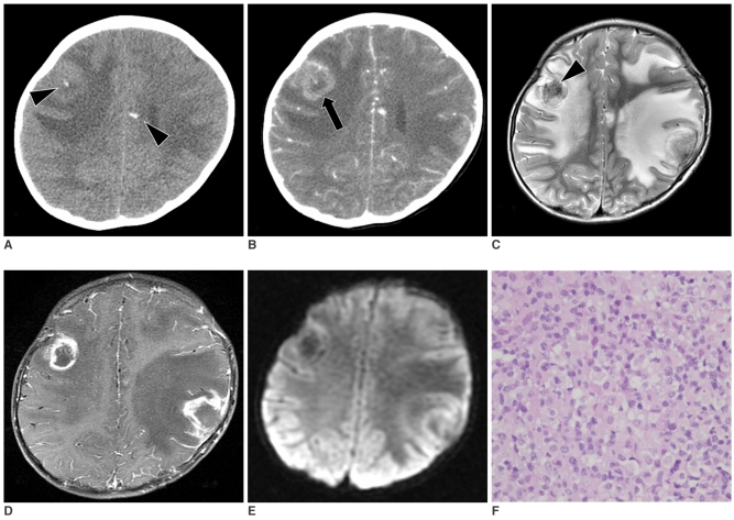
A 3-year-old boy with the hemophagocytic syndrome and who presented seizures and fever. A. On precontrast enhancement CT, several small parenchymal calcifications (arrowheads) can be seen in the subcortical area of both cerebral hemispheres. B. Contrast-enhanced CT reveals several thick walled peripheral enhancing lesions (arrow) with surrounding perilesional edema. Focal internal calcifications were also noted. C. The T2 weighted image reveals mixed signal intensity lesions with massive perilesional edema. The central, signal void portion of the right frontal mass shows calcification (arrowhead). D. The contrast enhanced T1 weighted image reveals irregular, thick, ring enhancement. E. No diffusion restriction in the lesions is noted on the diffusion weighted image. F. Many foamy histiocytes and atypical lymphocytes infiltrations are observed (H & E stain; original magnification, ×400).
Similar articles
-
A Diagnostic Dilemma: Similarity of Neuroradiological Findings in Central Nervous System Hemophagocytic Lymphohistiocytosis and Aspergillosis.Pediatr Blood Cancer. 2016 Jul;63(7):1296-9. doi: 10.1002/pbc.25967. Epub 2016 Mar 10. Pediatr Blood Cancer. 2016. PMID: 26970537
-
Frequency and development of CNS involvement in Chinese children with hemophagocytic lymphohistiocytosis.Pediatr Blood Cancer. 2010 Mar;54(3):408-15. doi: 10.1002/pbc.22239. Pediatr Blood Cancer. 2010. PMID: 19908295
-
Central nervous system involvement in adults with haemophagocytic lymphohistiocytosis: a single-center study.Ann Hematol. 2017 Aug;96(8):1279-1285. doi: 10.1007/s00277-017-3035-5. Epub 2017 Jun 7. Ann Hematol. 2017. PMID: 28589450
-
Familial hemophagocytic lymphohistiocytosis: clinical and neuroradiological findings and review of the literature.Childs Nerv Syst. 2010 Jan;26(1):121-7. doi: 10.1007/s00381-009-0957-9. Epub 2009 Aug 1. Childs Nerv Syst. 2010. PMID: 19649640 Review.
-
Hemophagocytic lymphohistiocytosis in the premature neonate.Adv Neonatal Care. 2009 Dec;9(6):265-73. doi: 10.1097/ANC.0b013e3181c20010. Adv Neonatal Care. 2009. PMID: 20010142 Review.
Cited by
-
Pediatric CNS-isolated hemophagocytic lymphohistiocytosis with brain hemorrhages: a case report.BMC Neurol. 2024 Oct 21;24(1):404. doi: 10.1186/s12883-024-03840-8. BMC Neurol. 2024. PMID: 39434014 Free PMC article.
-
Progressive Diffuse Osteonecrosis in a Patient with Secondary Hemophagocytic Lymphohistiocytosis.Case Rep Radiol. 2015;2015:730719. doi: 10.1155/2015/730719. Epub 2015 Nov 29. Case Rep Radiol. 2015. PMID: 26693376 Free PMC article.
-
Clinical, Genetic, and Outcome Characteristics of Pediatric Patients with Primary Hemophagocytic Lymphohistiocytosis.Turk Arch Pediatr. 2022 Jul;57(4):398-405. doi: 10.5152/TurkArchPediatr.2022.21314. Turk Arch Pediatr. 2022. PMID: 35822471 Free PMC article.
-
Autoimmune diseases of the brain, imaging and clinical review.Neuroradiol J. 2022 Apr;35(2):152-169. doi: 10.1177/19714009211042879. Epub 2021 Sep 7. Neuroradiol J. 2022. PMID: 34490814 Free PMC article. Review.
-
A spectrum of neuroradiological findings in children with haemophagocytic lymphohistiocytosis.Pediatr Radiol. 2007 Nov;37(11):1110-7. doi: 10.1007/s00247-007-0569-z. Epub 2007 Sep 5. Pediatr Radiol. 2007. PMID: 17846757
References
-
- Janka G, Imashuku S, Elinder G, Schneider M, Henter JI. Infection- and malignancy-associated hemophagocytic syndromes. Secondary hemophagocytic lymphohistiocytosis. Hematol Oncol Clin North Am. 1998;12:435–444. - PubMed
-
- Henter JI, Nennesmo I. Neuropathologic findings and neurologic symptoms in twenty-three children with hemophagocytic lymphohistiocytosis. J Pediatr. 1997;130:358–365. - PubMed
-
- Kieslich M, Vecchi M, Driever PH, Laverda AM, Schwabe D, Jacobi G. Acute encephalopathy as a primary manifestation of hemophagocytic lymphohistiocytosis. Dev Med Child Neurol. 2001;43:555–558. - PubMed
-
- Kollias SS, Ball WS, Jr, Tzika AA, Harris RE. Familial erythrophagocytic lymphohistiocytosis: neuroradiologic evaluation with pathologic correlation. Radiology. 1994;192:743–754. - PubMed
Publication types
MeSH terms
LinkOut - more resources
Full Text Sources
