

Carcinosarcoma of the liver with mesenchymal differentiation
- PMID: 17278210
- PMCID: PMC4066020
- DOI: 10.3748/wjg.v13.i5.809
Carcinosarcoma of the liver with mesenchymal differentiation
Abstract
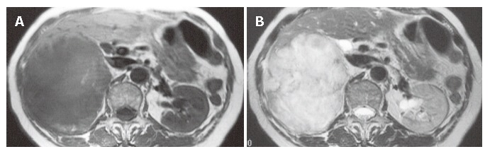
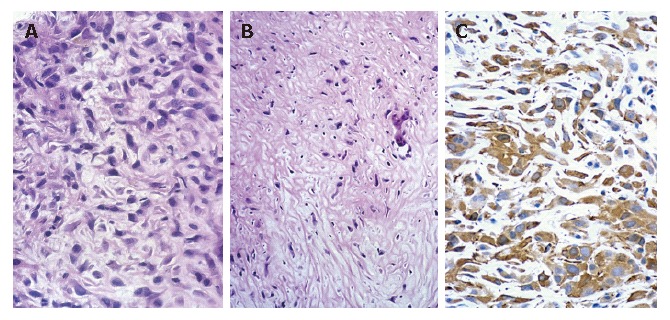
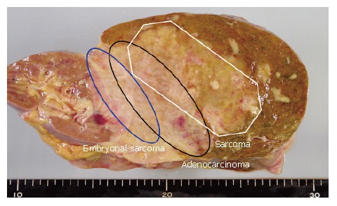
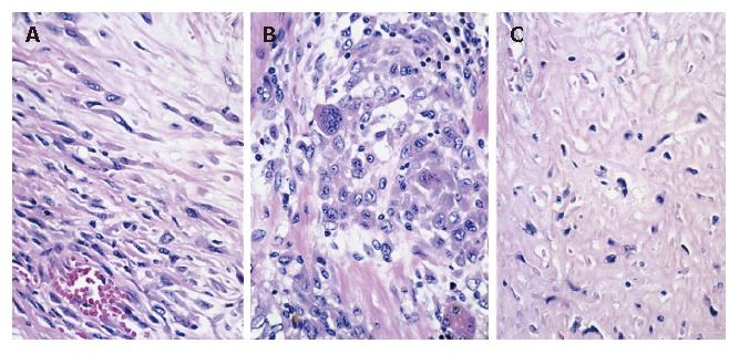
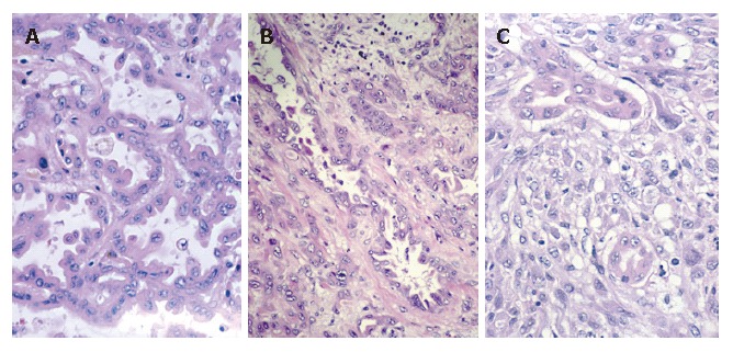
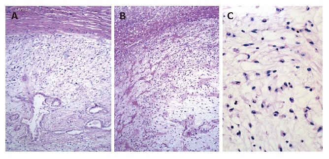
We report an extremely rare case where a mesenchymal differentiation, especially embryonal sarcoma, was demonstrated in cholangiocarcinoma. At autopsy, a yellowish-white tumor (15 cm x 12 cm) was found in the right hepatic lobe, and there were several daughter nodules in both hepatic lobes. Histologically, most of the main tumor and all of the daughter nodules examined showed sarcomatous changes (spindle cells, pleomorphic cells and hyalization). Histologic examination of a part of the main tumor disclosed a focus of adenocarcinoma within the tumor. The frequent transitions between the adenocarcinomatous areas and the sarcomatous areas suggested that sarcomatous transformation occurred in the cholangiocarcinoma and then spread rapidly. Immunohistochemically, the adenocarcinomatous elements were positive for cytokeratin, carcinoembryonic antigen (CEA) and epithelial membrane antigen, and negative in the sarcomatous cells. Vimentin was positive only in the sarcomatous elements. The findings of the present case support the view that carcinosarcomas represent carcinomas that develop sarcomatous elements via metaplasia of the epithelial element.
Figures
References
-
- Nakajima T, Kondo Y, Miyazaki M, Okui K. A histopathologic study of 102 cases of intrahepatic cholangiocarcinoma: histologic classification and modes of spreading. Hum Pathol. 1988;19:1228–1234. - PubMed
-
- Okuda K, Nakashima T. Primary carcinoma of the liver. 4th ed. In: Berk JE, editor. Bockus gastroenterology. Philadelphia: WB Saunders; 1985. pp. 3361–3364.
-
- Kakizoe S, Kojiro M, Nakashima T. Hepatocellular carcinoma with sarcomatous change. Clinicopathologic and immunohistochemical studies of 14 autopsy cases. Cancer. 1987;59:310–316. - PubMed
-
- Nakajima T, Kondo Y. A clinicopathologic study of intrahepatic cholangiocarcinoma containing a component of squamous cell carcinoma. Cancer. 1990;65:1401–1404. - PubMed
-
- Thompson L, Chang B, Barsky SH. Monoclonal origins of malignant mixed tumors (carcinosarcomas). Evidence for a divergent histogenesis. Am J Surg Pathol. 1996;20:277–285. - PubMed
Publication types
MeSH terms
LinkOut - more resources
Full Text Sources
Medical
