

NeuroTerrain--a client-server system for browsing 3D biomedical image data sets
- PMID: 17280615
- PMCID: PMC1802997
- DOI: 10.1186/1471-2105-8-40
NeuroTerrain--a client-server system for browsing 3D biomedical image data sets
Abstract
Background: Three dimensional biomedical image sets are becoming ubiquitous, along with the canonical atlases providing the necessary spatial context for analysis. To make full use of these 3D image sets, one must be able to present views for 2D display, either surface renderings or 2D cross-sections through the data. Typical display software is limited to presentations along one of the three orthogonal anatomical axes (coronal, horizontal, or sagittal). However, data sets precisely oriented along the major axes are rare. To make fullest use of these datasets, one must reasonably match the atlas' orientation; this involves resampling the atlas in planes matched to the data set. Traditionally, this requires the atlas and browser reside on the user's desktop; unfortunately, in addition to being monolithic programs, these tools often require substantial local resources. In this article, we describe a network-capable, client-server framework to slice and visualize 3D atlases at off-axis angles, along with an open client architecture and development kit to support integration into complex data analysis environments.
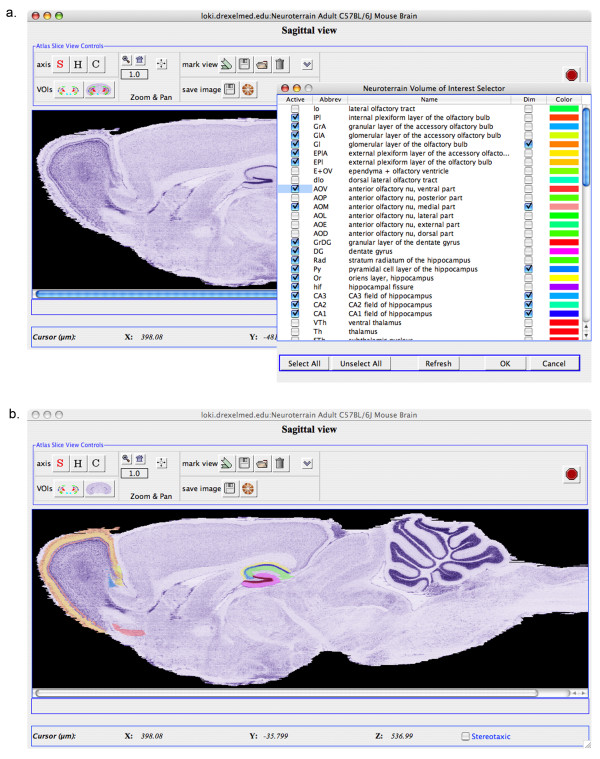
Results: Here we describe the basic architecture of a client-server 3D visualization system, consisting of a thin Java client built on a development kit, and a computationally robust, high-performance server written in ANSI C++. The Java client components (NetOStat) support arbitrary-angle viewing and run on readily available desktop computers running Mac OS X, Windows XP, or Linux as a downloadable Java Application. Using the NeuroTerrain Software Development Kit (NT-SDK), sophisticated atlas browsing can be added to any Java-compatible application requiring as little as 50 lines of Java glue code, thus making it eminently re-useable and much more accessible to programmers building more complex, biomedical data analysis tools. The NT-SDK separates the interactive GUI components from the server control and monitoring, so as to support development of non-interactive applications. The server implementation takes full advantage of data center's high-performance hardware, where it can be co-localized with centrally-located, 3D dataset repositories, extending access to the researcher community throughout the Internet.
Conclusion: The combination of an optimized server and modular, platform-independent client provides an ideal environment for viewing complex 3D biomedical datasets, taking full advantage of high-performance servers to prepare images and subsets of associated meta-data for viewing, as well as the graphical capabilities in Java to actually display the data.
Figures


Similar articles
-
JAtlasView: a Java atlas-viewer for browsing biomedical 3D images and atlases.BMC Bioinformatics. 2005 Mar 9;6:47. doi: 10.1186/1471-2105-6-47. BMC Bioinformatics. 2005. PMID: 15757508 Free PMC article.
-
Web tools for large-scale 3D biological images and atlases.BMC Bioinformatics. 2012 Jun 7;13:122. doi: 10.1186/1471-2105-13-122. BMC Bioinformatics. 2012. PMID: 22676296 Free PMC article.
-
The design of Jemboss: a graphical user interface to EMBOSS.Bioinformatics. 2003 Sep 22;19(14):1837-43. doi: 10.1093/bioinformatics/btg251. Bioinformatics. 2003. PMID: 14512356
-
PET/CT image navigation and communication.J Nucl Med. 2004 Jan;45 Suppl 1:46S-55S. J Nucl Med. 2004. PMID: 14736835 Review.
-
Visualization approaches for multidimensional biological image data.Biotechniques. 2007 Jul;43(1 Suppl):31, 33-6. doi: 10.2144/000112511. Biotechniques. 2007. PMID: 17936940 Review.
Cited by
-
Visualizing data mining results with the brede tools.Front Neuroinform. 2009 Jul 28;3:26. doi: 10.3389/neuro.11.026.2009. eCollection 2009. Front Neuroinform. 2009. PMID: 19668704 Free PMC article.
-
3D brain atlas reconstructor service--online repository of three-dimensional models of brain structures.Neuroinformatics. 2013 Oct;11(4):507-18. doi: 10.1007/s12021-013-9199-9. Neuroinformatics. 2013. PMID: 23943281 Free PMC article.
-
neuroVIISAS: approaching multiscale simulation of the rat connectome.Neuroinformatics. 2012 Jul;10(3):243-67. doi: 10.1007/s12021-012-9141-6. Neuroinformatics. 2012. PMID: 22350719
-
MBAT: a scalable informatics system for unifying digital atlasing workflows.BMC Bioinformatics. 2010 Dec 22;11:608. doi: 10.1186/1471-2105-11-608. BMC Bioinformatics. 2010. PMID: 21176225 Free PMC article.
-
A proposal for a coordinated effort for the determination of brainwide neuroanatomical connectivity in model organisms at a mesoscopic scale.PLoS Comput Biol. 2009 Mar;5(3):e1000334. doi: 10.1371/journal.pcbi.1000334. Epub 2009 Mar 27. PLoS Comput Biol. 2009. PMID: 19325892 Free PMC article.
References
-
- Rosen GD, La Porte NT, Diechtiareff B, Pung CJ, Nissanov J, Gustafson C, Bertrand L, Gefen S, Fan Y, Tretiak OJ, Manly KF, Park MR, Williams AG, Connolly MT, Capra JA, Williams RW. Informatics center for mouse genomics: the dissection of complex traits of the nervous system. Neuroinformatics. 2003;1:327–342. doi: 10.1385/NI:1:4:327. - DOI - PubMed
-
- Bug W, Gustafson C, Shahar A, Gefen S, Fan Y, Bertrand L, Nissanov J. Brain spatial normalization: indexing neuroanatomical databases. In: Chaquito C, editor. Neuroinformatics, in press. Totowa, NJ, USA , Humana Press; 2006. (Methods in Molecular Biology). Walker J. - PubMed
Publication types
MeSH terms
Grants and funding
LinkOut - more resources
Full Text Sources
Medical
Research Materials
