. 2007 Apr;143(4):607-15.
doi: 10.1016/j.ajo.2006.12.006. Epub 2007 Feb 5.

Changes in select redox proteins of the retinal pigment epithelium in age-related macular degeneration

Affiliations

Changes in select redox proteins of the retinal pigment epithelium in age-related macular degeneration

Alejandra Decanini et al. Am J Ophthalmol. 2007 Apr.

Abstract

Purpose: To examine changes of select reduction-oxidation (redox) sensitive proteins from human donor retinal pigment epithelium (RPE) at four stages of age-related macular degeneration (AMD).

Design: Experimental study.

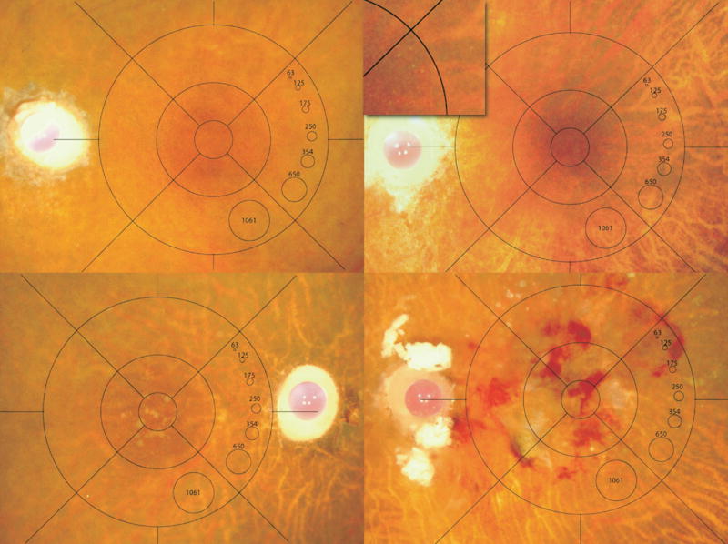
Methods: Human donor eyes were obtained from the Minnesota Lions Eye Bank and graded using the Minnesota Grading System (MGS) into four stages that correspond to stages defined by the age-related eye disease study (AREDS). Protein content in RPE homogenates was measured using Western immunoblotting with protein-specific antibodies.

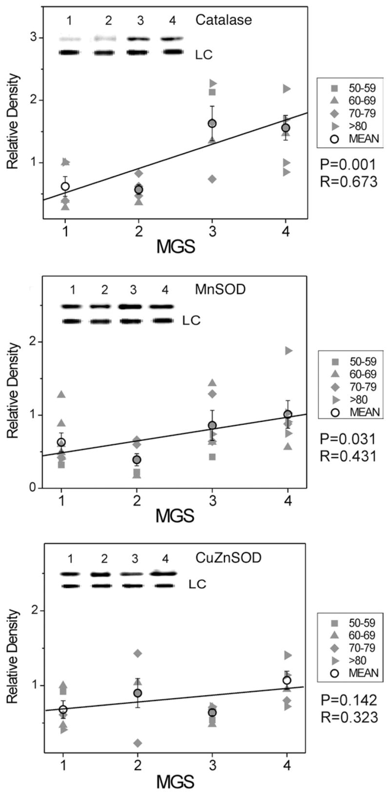
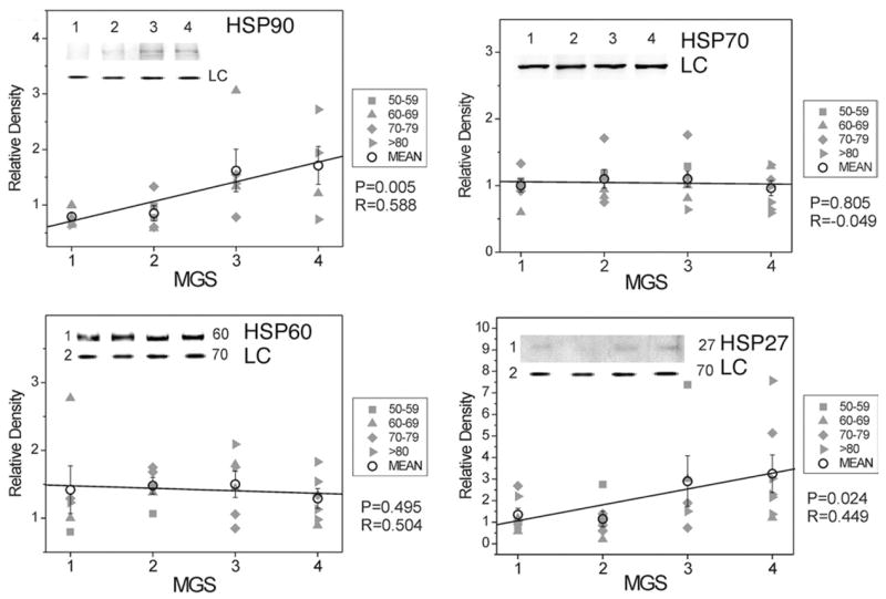
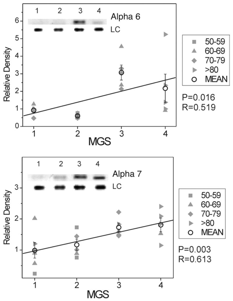
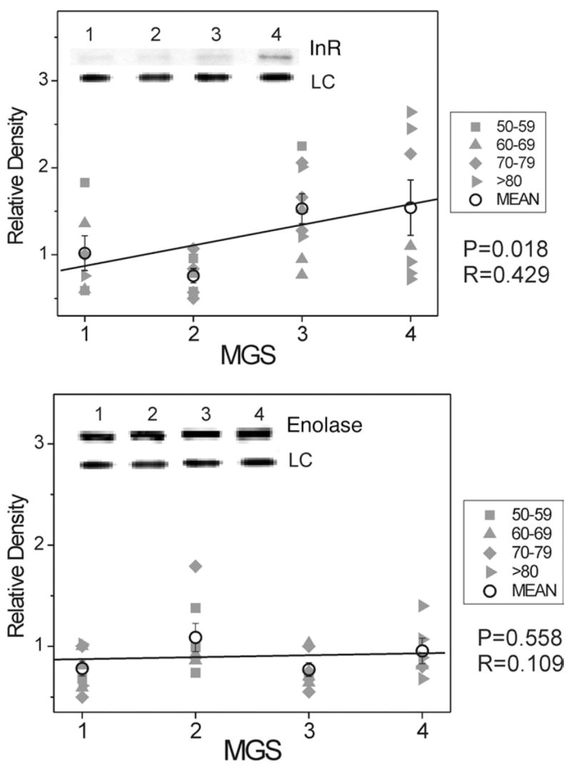
Results: The content of several antioxidant enzymes and specific proteins that facilitate refolding or degradation of oxidatively damaged proteins increased significantly in MGS stage 3. These proteins are involved in the primary (copper-zinc superoxide dismutase [CuZnSOD], manganese superoxide dismutase [MnSOD], and catalase) and secondary (heat shock protein [HSP] 27, HSP 90, and proteasome) defense against oxidative damage. Additionally, the insulin pro-survival receptor exhibited disease-related upregulation.

Conclusions: The pattern of protein changes identified in human donor tissue graded using the MGS support the role of oxidative mechanisms in the pathogenesis and progression of AMD. The MGS uses nearly identical clinical definitions and grading criteria of AMD that are used in the AREDS, so our results apply to clinical and epidemiologic studies using similar definitions. Results from our protein analysis of human donor tissue helps to explain altered oxidative stress regulation and cell-survival pathways that occur in progressive stages of AMD.


Figures

FIGURE 1
FIGURE 1
Minnesota Grading System (MGS) stages 1–4. Representative, digital images (stereo not shown) of human eye bank eyes for each stage that corresponds to the clinical definitions used in the Age-Related Eye Disease Study (AREDS). Images demonstrate MGS1 (Top left), MGS2 (Top right; inset showing small hard drusen), MGS3 (Bottom left), and MGS4 (Bottom right).
FIGURE 2
FIGURE 2
Select redox proteins in Minnesota Grading System (MGS) stages of age-related macular degeneration (AMD); antioxidant enzyme content. Retinal pigment epithelial (RPE) proteins (15 μg) were subjected to sodium dodecyl sulphate polyacrylamide gel electrophoresis followed by Western immunoblotting. Data were normalized to a standard reaction in all blots and to heat shock protein 70 (HSP70) as a loading control (LC). Individual donor relative densities from four age groups are shown (gray symbols). Group mean ± standard error of the mean (SEM) and the results of linear regression analysis are shown. n = 5–7 donors/stage. Inset shows representative Western immunoblot of antioxidant enzymes catalase (Top), manganese superoxide dismutase (MnSOD) (Middle), and copper-zinc superoxide dismutase (CuZnSOD) (Bottom) at MGS 1–4 and the LC-HSP70.
FIGURE 3
FIGURE 3
Select redox proteins in Minnesota Grading System (MGS) stages of age-related macular degeneration (AMD); heat shock protein (HSP) content. Retinal pigment epithelial (RPE) proteins (15 μg) were subjected to sodium dodecyl sulphate polyacrylamide gel electrophoresis followed by Western immunoblotting. Data were normalized to a standard reaction in all blots and to HSP70 as a loading control (LC). Individual donor relative densities from four age groups are shown (gray symbols). Group mean ± standard error of the mean (SEM) and the results of linear regression analysis are shown. n = 5–7 donors/stage. Inset shows representative Western immunoblot of heat shock proteins HSP90 (Top left), HSP70 (Top right), HSP60 (Bottom left), and HSP27 (Bottom right) at MGS 1–4 and the LC.
FIGURE 4
FIGURE 4
Select redox proteins in Minnesota Grading System (MGS) stages of age-related macular degeneration (AMD); total proteasome content. Retinal pigment epithelial (RPE) proteins (15 μg) were subjected to sodium dodecyl sulphate polyacrylamide gel electrophoresis followed by Western immunoblotting. Data were normalized to a standard reaction in all blots and to heat shock protein 70 (HSP70) as a loading control (LC). Individual donor relative densities from four age groups are shown (gray symbols). Group mean ± standard error of the mean (SEM) and the results of linear regression analysis are shown. n = 5–7 donors/stage. Inset shows representative Western immunoblot for proteasome subunits Alpha 6 (Top) and Alpha 7 (Bottom) at MGS 1–4 and the LC.
FIGURE 5
FIGURE 5
Select redox proteins in Minnesota Grading System (MGS) stages of age-related macular degeneration (AMD); insulin receptor and enolase content. Retinal pigment epithelial (RPE) proteins (15 μg) were subjected to sodium dodecyl sulphate polyacrylamide gel electrophoresis followed by Western immunoblotting. Data were normalized to a standard reaction in all blots and to heat shock protein 70 (HSP70) as a loading control (LC). Individual donor relative densities from four age groups are shown (gray symbols). Group mean ± standard error of the mean (SEM) and the results of linear regression analysis are shown. n = 5–9 donors/stage. Inset shows representative Western immunoblot for insulin receptor β subunit (Top) and enolase (Bottom) at MGS 1–4 and the LC.

References

    1. Leibowitz HM, Krueger DE, Maunder LR, et al. The Framingham Eye Study monograph: an ophthalmological and epidemiological study of cataract, glaucoma, diabetic retinopathy, macular degeneration, and visual acuity in a general population of 2631 adults, 1973–1975. Surv Ophthalmol. 1980;24:335–610. - PubMed
    1. Klein BE, Klein R. Cataracts and macular degeneration in older Americans. Arch Ophthalmol. 1982;100:571–573. - PubMed
    1. Hyman LG, Lilienfeld AM, Ferris FL, Fine SL. Senile macular degeneration: a case-control study. Am J Epidemiol. 1983;118:213–227. - PubMed
    1. Klein R, Klein BE, Linton KL. Prevalence of age-related maculopathy: the Beaver Dam Eye Study. Ophthalmology. 1992;99:933–943. - PubMed
    1. Klein R, Wang Q, Klein BE, et al. The relationship of age-related maculopathy, cataract, and glaucoma to visual acuity. Invest Ophthalmol Vis Sci. 1995;36:182–191. - PubMed

Publication types

MeSH terms