Review
. 2008 Jan 12;363(1489):57-70.
doi: 10.1098/rstb.2006.2012.

Regulatory pathways linking progenitor patterning, cell fates and neurogenesis in the ventral neural tube

Affiliations
Review

Regulatory pathways linking progenitor patterning, cell fates and neurogenesis in the ventral neural tube

James Briscoe et al. Philos Trans R Soc Lond B Biol Sci. .

Abstract

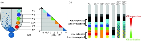
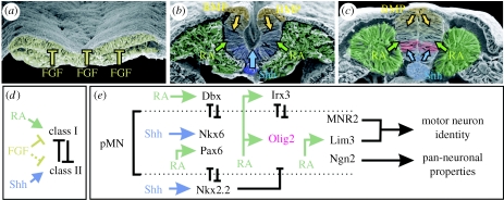
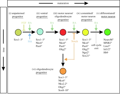
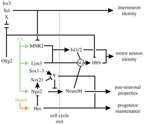
The assembly of neural circuits in the vertebrate central nervous system depends on the organized generation of specific neuronal subtypes. Studies over recent years have begun to reveal the principles and elucidate some of the detailed mechanisms that underlie these processes. In general, exposure to different types and concentrations of signals directs neural progenitor populations to generate specific subtypes of neurons. These signals function by regulating the expression of intrinsic determinants, notably transcription factors, which specify the fate of cells as they differentiate into neurons. In this review, we illustrate these concepts by focusing on the generation of neurons in ventral regions of the spinal cord, where detailed knowledge of the mechanisms that regulate cell identity has provided insight into the development of a number of neuronal subtypes, including motor neurons. A greater knowledge of the molecular control of neural development is likely to have practical benefits in understanding the causes and consequences of neurological diseases. Moreover, recent studies have demonstrated how an understanding of normal neural development can be applied to direct differentiation of stem cells in vitro to specific neuronal subtypes. This type of rational manipulation of stem cells may represent the first step in the development of treatments based on therapeutic replacement of diseased or damaged nervous tissue.


Figures

Figure 1
Figure 1
The generation of neuronal subtypes in the ventral spinal cord by a gradient of Shh/Gli signalling. Distinct neuronal subtypes are generated along the dorsoventral axis in the ventral neural tube in response to graded Shh signalling. (a) Five distinct ventral neuronal subtypes arise from an equivalent number of progenitor domains in the ventricular zone of the ventral spinal cord. Progressively, more dorsal progenitor domains are exposed to a decreasing concentration of Shh protein and, in vitro, the concentration of Shh determines the neuronal subtype generated. (b) Dorsoventral spinal cord patterning defects in Gli mutant embryos can be explained by a gradient model of Gli activator and repressor activity. The diagrams illustrate the effects of Shh and Gli gene loss-of-function mutations on the patterning of the ventral and intermediate regions of the spinal cord. Gli1−/− mutants have no defect in ventral patterning. In Gli2−/− embryos, the most ventral cell types, the FP and V3 neurons, are absent and there is a concomitant expansion of the MN domain. By contrast, in Gli3−/− mutants, patterning of the intermediate region of the neural tube is disrupted. Embryos lacking both Shh and Gli3 functions demonstrate a substantial restoration in normal dorsoventral patterning, compared to Shh null mutants alone and a similar situation is observed in Gli2−/− and Gli3−/− mutants.
Figure 2
Figure 2
Signalling pathways leading to motor neuron generation. Scanning electron micrographs of the developing chick neural tube at (a) neural plate, (b) neural folds and (c) neural tube stages (images generously provided by Dr Kathryn Tosney, University of Michigan). Images display sources of FGF (yellow), retinoic acid (RA, green), Shh (blue) and BMPs (orange) surrounding the neural tissue and influencing progenitor gene expression. (d) Influence of FGF, RA and Shh on class I and II protein expressions. FGF signalling prevents the emergence of dorsal–ventral pattern by blocking both class I and II protein expressions. RA signalling induces the expression of class I proteins, whereas Shh signalling appears to induce class II proteins. Once expressed, reciprocal pairs of class I and II proteins exhibit cross-repressive interactions to partition the ventral neural tube into discrete progenitor domains. (e) Shh and RA initiate the expression of the HD proteins Nkx6.1, Nkx6.2 (Nkx6) and Pax6 in ventral spinal cord progenitors. The repressor activities of Nkx6 and Pax6 prevent the expression of inhibitors of MN formation, such as Dbx1, Dbx2 (Dbx) and Nkx2.2, and permit ligand-bound retinoid receptors to activate Olig2 expression (red). Within MN progenitors, Olig2 directs MN differentiation by repressing Irx3 and other unidentified target genes that regulate the expression of the MN-specific transcription factors, MNR2, Lim3, Isl1, Isl2 and Hb9, and the pan-neuronal transcription factors, Ngn2 and NeuroM/Math3. This repressor activity of Olig2 works in conjunction with the activator function of ligand-bound retinoid receptors.
Figure 3
Figure 3
Progressive changes in progenitor cells during motor neuron development. (i) Neural progenitors expressing Sox1–3 are held in an undifferentiated and unpatterned state through the actions of FGFs. (ii) The combined actions of Shh and RA signalling ventralizes neural progenitors through the induction of progenitor transcription factors, such as Nkx6. (iii) Continued exposure to Shh and RA leads to the induction of Olig2 in bipotential motor neuron/oligodendrocyte progenitors. (iv) In the presence of RA and absence of Notch signalling, Olig2+ cells express Ngn2 and Sox21 and begin to exit the cell cycle and differentiate into motor neurons. (v) The further differentiation of motor neurons requires the ongoing RA signalling, as well as the downregulation of Olig2. (vi) Activation of the Notch signalling pathway prevents the expression of Ngn2 in Olig2+ motor neuron/oligodendrocyte progenitors and maintains these cells in an undifferentiated state until additional temporal cues direct the formation of oligodendrocytes from these cells. Notch signalling may also positively promote oligodendrocyte development (dashed line). See text for more details.
Figure 4
Figure 4
Olig2 and the transcription factor network controlling motor neuron differentiation. Olig2 promotes motor neuron development through its ability to repress the expression of antagonists of motor neuron formation, such as Irx3, Scl and other unidentified target genes (X). The actions of ligand-bound retinoic acid receptors lead to the expression of transcription factors that act downstream of Olig2, including MNR2, Lim3 and Ngn2. The expression of Ngn2 is negatively regulated by Hes proteins that act downstream of the Notch signalling pathway. The balance of SoxB activator and repressor functions within motor neuron progenitors further regulates the activity of Ngn2 (Bylund et al. 2003; Sandberg et al. 2005). The activator properties of Sox1–3 appear to induce the expression of unknown inhibitors of Ngn2 function (Y). Sox21 acts downstream of Ngn2 and opposes the actions of Sox1–3 by reducing the expression of Y, thereby elevating Ngn2 activity and facilitating motor neuron differentiation. Through their ability to block Ngn2 expression and activity, Notch signalling, Hes genes and Sox1–3 maintain progenitors in an undifferentiated state. The release of Ngn2 from these regulatory constraints leads to the upregulation of NeuroM and the exit of progenitors from the cell cycle. NeuroM then forms a transcriptional activator complex with Lim3 and Isl1/2, mediated by NLI, that displaces an Olig2 repressor complex from the Hb9 promoter and activates Hb9 expression to drive motor neuron-specific gene expression (Lee et al. 2005).

Similar articles

Cited by

References

    1. Altman J, Bayer S.A. The development of the rat spinal cord. Adv. Anat. Embryol. Cell Biol. 1984;85:1–164. - PubMed
    1. Aza-Blanc P, Lin H, Ruiz I.A.A, Kornberg T.B. Expression of the vertebrate gli proteins in Drosophila reveals a distribution of activator and repressor activities. Development. 2000;127:4293–4301. - PubMed
    1. Bai C.B, Joyner A.L. Gli1 can rescue the in vivo function of Gli2. Development. 2001;128:5161–5172. - PubMed
    1. Bai C.B, Stephen D, Joyner A.L. All mouse ventral spinal cord patterning by Hedgehog is Gli dependent and involves an activator function of Gli3. Dev. Cell. 2004;6:103–115. doi:10.1016/S1534-5807(03)00394-0 - DOI - PubMed
    1. Barth K.A, Kishimoto Y, Rohr K.B, Seydler C, Schulte-Merker S, Wilson S.W. Bmp activity establishes a gradient of positional information throughout the entire neural plate. Development. 1999;126:4977–4987. - PubMed

Publication types