. 2007 Apr;27(8):3176-86.
doi: 10.1128/MCB.01652-06. Epub 2007 Feb 5.

Disruption of the FEN-1/PCNA interaction results in DNA replication defects, pulmonary hypoplasia, pancytopenia, and newborn lethality in mice

Affiliations

Disruption of the FEN-1/PCNA interaction results in DNA replication defects, pulmonary hypoplasia, pancytopenia, and newborn lethality in mice

Li Zheng et al. Mol Cell Biol. 2007 Apr.

Abstract

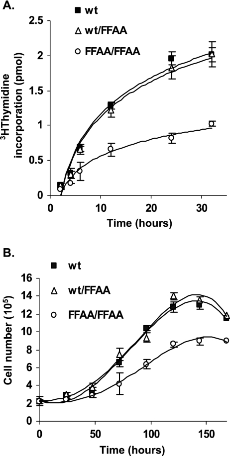
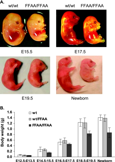
The interaction between flap endonuclease 1 (FEN-1) and proliferation cell nuclear antigen (PCNA) is critical for faithful and efficient Okazaki fragment maturation. In a living cell, this interaction is probably important for PCNA to load FEN-1 to the replication fork, to coordinate the sequential functions of FEN-1 and other enzymes, and to stimulate its enzyme activity. The FEN-1/PCNA interaction is mediated by the motif (337)QGRLDDFFK(345) of FEN-1, such that an F343AF344A (FFAA) mutant cannot bind to PCNA but retains its nuclease activities. To determine the physiological roles of the FEN-1/PCNA interaction in a mammalian system, we knocked the FFAA Fen1 mutation into the Fen1 gene locus of mice. FFAA/FFAA mouse embryo fibroblasts underwent DNA replication and division at a slower pace, and FFAA/FFAA mutant embryos displayed significant defects in growth and development, particularly in the lung and blood systems. All newborn FFAA mutant pups died at birth, likely due to pulmonary hypoplasia and pancytopenia. Collectively, our data demonstrate the importance of the FEN-1/PCNA complex in DNA replication and in the embryonic development of mice.


Figures

FIG. 1.
FIG. 1.
Generation of F343AF344A (FFAA) knock-in mice. (A) Expected restriction maps of FFAA targeting vector and different types of mouse Fen1 alleles. (B) Southern hybridization analysis of wt and FFAA mutant (neo+) alleles. Genomic DNA isolated from wt and FFAA (neo+) ES cells was digested with HindIII (upper panel) or XhoI (lower panel) and detected with probe 1 or 2, respectively. DNA sequences of probes 1 and 2 correspond to the sequences from 19185 to 19673 and 5155 to 5640, respectively, of BAC RP22-325J22, chromosome 19 of Mus musculus (129S1). Probes 1 and 2 were prepared by PCR amplification using primers listed in Table 1. Cleavage of wild-type genomic DNA by HindIII or XhoI generated 8.7-kb and 13.1-kb bands, respectively. Digestion of genomic DNA of FFAA (neo+) by HindIII produced two 8.7-kb and 5.8-kb bands, and digestion of the genomic DNA by XhoI produced two 13.1-kb and 10.5-kb bands. (C) Genotype determination by PCR. Fen1 alleles were amplified by PCR using primers mFEN125 and mFEN166 (Table 1) and mouse tail genomic DNA as a template. PCR products were resolved in a 2% agarose gel. PCR product amplified from wt/wt genomic DNA gave a single band of 1,066 bp, whereas the PCR product from FFAA/FFAA genomic DNA gave a single band of 1,121 bp due to the existence of a copy of Lox P sequence. Thus, the PCR product from wt/FFAA genomic DNA gave two bands of 1,066 and 1,121 bp, respectively. The upper panel shows a schematic of the assay, and the bottom panel shows the gel image of a genotype analysis of six mice. Lanes 1 and 4 indicate the genotype is wt. Lanes 2, 3, and 6 indicate the genotype is heterozygous, and lane 5 indicates the genotype is homozygous. (D) Sequence confirmation of mice homozygous for FFAA. Fen1 alleles were amplified by PCR using tail genomic DNA as a template and mFFAA-1 and mFFAA-4 as primers (Table 1). The DNA sequence encoding the 343 and 344 amino acid residues of different mFEN-1 alleles was confirmed by direct DNA sequencing using primer mFFAA-3 (Table 1).
FIG. 2.
FIG. 2.
The FFAA mutation eliminates FEN-1/PCNA interaction and impairs the recruitment of FEN-1 to replication foci. (A) Mouse recombinant FFAA FEN-1 mutant is unable to interact with mouse PCNA. His6-tagged FEN-1 or nontagged PCNA was expressed in E. coli. Cell lysates containing recombinant His6-tagged FEN-1 or His6-tagged FFAA were mixed with the lysate of nontagged PCNA. The FEN-1 or FFAA was purified with Ni2+ chelating agarose. The PCNA in complex with FEN-1 was analyzed with 4 to 15% SDS-PAGE. (B) Coimmunoprecipitation of FEN-1 and PCNA. Whole-cell extract (CE) was prepared from wt or FFAA/FFAA MEFs. The wt or FFAA/FFAA CE was incubated with monoclonal anti-FEN-1 antibody-conjugated protein G beads. After an extensive wash, proteins bound to protein G beads were analyzed by Western blotting using polyclonal anti-PCNA antibody or monoclonal anti-FEN-1 antibody. (C) Representative images of localization of FEN-1 and PCNA in cells at the G1/S boundary and cells in S phase. wt and FFAA/FFAA MEFs were synchronized at the G1/S boundary by mimosine treatment. Upon removal of mimosine, cells were released into S phase. Typically, 60% of cells were in S phase at 4 h post-mimosine treatment. Cells were fixed and costained with anti-FEN-1 and anti-PCNA antibodies. In the merge views, yellow spots indicate colocalization of FEN-1 and PCNA. (D) Representative images of localization of FEN-1 and BrdU in cells at the G1/S boundary and cells in S phase. wt and FFAA/FFAA MEFs at the G1/S boundary or in S phase were labeled with BrdU for 1 h. Cells were fixed and costained with anti-FEN-1 and anti-BrdU antibodies.
FIG. 3.
FIG. 3.
FFAA/FFAA nuclear extracts are deficient in Okazaki fragment maturation (OFM) in vitro. Four synthetic substrates that mimic various OFM intermediates were utilized in a nick translation assay to compare the OFM efficiency of nuclear extracts generated from wt with those from homozygous FFAA mouse cells. As a control, wt nuclear extracts that were FEN-1 immunodepleted (ΔFEN-1) were assayed as well. Nick translation on gap substrates (A) R3, (B) R1, and (C) R0 having three, one, and zero 5′ ribonucleotides, respectively. (D) Nick translation on a gap substrate with a 5-nt 5′ flap (F5). In each panel, a schematic of the substrate used, a representative gel image from the experiment, and a bar graph depicting the quantitation of the product band are shown. Oligonucleotides used to make R3, R1, R0, and F5 are listed in Table 1.
FIG. 4.
FIG. 4.
FFAA/FFAA cells have retarded DNA replication and cell proliferation. (A) The DNA replication efficiency in wt, wt/FFAA, and FFAA/FFAA MEFs was measured by 3H incorporation assay. Cells were incubated with [3H]thymidine for 2, 4, 6, 12, or 24 h. The 3H activity that was incorporated into genomic DNA was determined by a liquid scintillation counter. Values are averages of at least five independent assays. (B) The cell proliferation rate of wt, wt/FFAA, and FFAA/FFAA MEFs. Each value represents the average of three independent measurements of independent embryos for each genetic background.
FIG. 5.
FIG. 5.
Retarded growth and defective development of FFAA/FFAA embryos. (A) Gross images of wt and FFAA/FFAA embryos from E14.5 to E19.5. The FFAA/FFAA embryos are visibly smaller than the wt littermates. (B) Weight comparison of wt, wt/FFAA, and FFAA/FFAA embryos. Values are means ± standard deviations of the weight of at least 10 embryos for each category.
FIG. 6.
FIG. 6.
FFAA/FFAA embryos develop pulmonary hypoplasia. Anatomical and histological analyses were performed on wt, wt/FFAA, and FFAA/FFAA embryos at E19.5. Because wt and wt/FFAA embryos exhibited identical phenotypes, the images of wt/FFAA embryos were not displayed. (A) Macrographic images of the lungs of a wt and an FFAA/FFAA embryo. (B) Lung/body ratios of the wild-type and FFAA mouse embryos. Values are means ± standard deviations of measurements from at least 10 embryos on each genetic background. (C and D) Micrographic images (×100) and higher-magnification views (insets, ×400) of the lung of a wt (C) and an FFAA/FFAA (D) embryo (H&E stain).
FIG. 7.
FIG. 7.
Impairment of embryonic hematopoiesis and peripheral pancytopenia. (A and B) Histological features of embryonic hematopoiesis in the liver of a wt (A) and an FFAA/FFAA embryo (B) at E19.5 showing liver extramedullary hematopoiesis (H&E stain; ×200). The FFAA/FFAA embryo displayed markedly impaired hematopoiesis. (C and D) Blood smears of a wt (C) and an FFAA/FFAA embryo (D) at E19.5. Blood smears were prepared using blood sampling from the corresponding embryo hearts (Wright-Giemsa stain; ×200).

Similar articles

Cited by

References

    1. Adachi, N., Z. E. Karanjawala, Y. Matsuzaki, H. Koyama, and M. R. Lieber. 2002. Two overlapping divergent transcription units in the human genome: the FEN1/C11orf10 locus. OMICS J. Integr. Biol. 6:273-279. - PubMed
    1. Ayyagari, R., X. V. Gomes, D. A. Gordenin, and P. M. Burgers. 2003. Okazaki fragment maturation in yeast. I. Distribution of functions between FEN1 and DNA2. J. Biol. Chem. 278:1618-1625. - PubMed
    1. Bae, S. H., K. H. Bae, J. A. Kim, and Y. S. Seo. 2001. RPA governs endonuclease switching during processing of Okazaki fragments in eukaryotes. Nature 412:456-461. - PubMed
    1. Biswas, E. E., F. X. Zhu, and S. B. Biswas. 1997. Stimulation of RTH1 nuclease of the yeast Saccharomyces cerevisiae by replication protein A. Biochemistry 36:5955-5962. - PubMed
    1. Bravo, R., and H. Macdonald-Bravo. 1987. Existence of two populations of cyclin/proliferating cell nuclear antigen during the cell cycle: association with DNA replication sites. J. Cell Biol. 105:1549-1554. - PMC - PubMed

Publication types

MeSH terms

Substances