. 2007 Apr;27(8):3056-64.
doi: 10.1128/MCB.01339-06. Epub 2007 Feb 5.

Rbm15 modulates Notch-induced transcriptional activation and affects myeloid differentiation

Affiliations

Rbm15 modulates Notch-induced transcriptional activation and affects myeloid differentiation

Xianyong Ma et al. Mol Cell Biol. 2007 Apr.

Abstract

RBM15 is the fusion partner with MKL in the t(1;22) translocation of acute megakaryoblastic leukemia. To understand the role of the RBM15-MKL1 fusion protein in leukemia, we must understand the normal functions of RBM15 and MKL. Here, we show a role for Rbm15 in myelopoiesis. Rbm15 is expressed at highest levels in hematopoietic stem cells and at more moderate levels during myelopoiesis of murine cell lines and primary murine cells. Decreasing Rbm15 levels with RNA interference enhances differentiation of the 32DWT18 myeloid precursor cell line. Conversely, enforced expression of Rbm15 inhibits 32DWT18 differentiation. We show that Rbm15 alters Notch-induced HES1 promoter activity in a cell type-specific manner. Rbm15 inhibits Notch-induced HES1 transcription in nonhematopoietic cells but stimulates this activity in hematopoietic cell lines, including 32DWT18 and human erythroleukemia cells. Moreover, the N terminus of Rbm15 coimmunoprecipitates with RBPJkappa, a critical factor in Notch signaling, and the Rbm15 N terminus has a dominant negative effect, impairing activation of HES1 promoter activity by full-length-Rbm15. Thus, Rbm15 is differentially expressed during hematopoiesis and may act to inhibit myeloid differentiation in hematopoietic cells via a mechanism that is mediated by stimulation of Notch signaling via RBPJkappa.


Figures

FIG. 1.
FIG. 1.
Mouse RBM15 protein sequence and subcellular localization. (A) Full-length mouse Rbm15 protein sequence, with predicted nuclear localization signals shown in boldface. (B) CHO cells were transfected with plasmids encoding GFP (left) or GFP-Rbm15 (right) proteins. The upper panels show Rbm15 subcellular localization (right) (based on GFP fluorescence) by confocal microscopy at 24 h posttransfection. Images in the lower panels reveal the autofluorescence of the cells above. (C) Western blot analysis of CHO cells transiently transfected with plasmid encoding GFP or GFP-Rbm15. Cytoplasmic extract (CE) and nuclear extract (NE) were derived from the CHO cells, and the transgenes (as indicated above each lane) were detected with anti-GFP. Numbers at right represent relative locations of molecular mass standards.
FIG. 2.
FIG. 2.
Rbm15 expression in tissues and myeloid cells. (A to C) Analysis of Rbm15 using a murine multitissue Northern blot (A), as well as during myeloid differentiation of EML (B) and 32DWT18 cells (C). β-Actin mRNA was probed as a loading control (bottom). Note that two forms of Rbm15 predominate, one at approximately 9 kb (full length [FL]) and the other at 4 kb (spliced [SP]) in the tissues but not in the myeloid cell lines. (D) qRT-PCR data for Rbm15 in primary lineage-negative murine bone marrow cells, as well as in macrophages and megakaryocytes differentiated from primary bone marrow. Error bars indicate standard deviations.
FIG. 3.
FIG. 3.
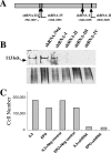
Targeted inhibition of Rbm15 expression by RNA interference. (A) Schematic showing locations of four shRNAs tested for inhibition of Rbm15 expression. (B) Transient cotransfection of CHO cells using a GFP-Rbm15 expression plasmid together with shRNA-I, shRNA-II, shRNA-III, shRNA-IV, or negative control (luciferase shRNA) retroviral constructs separately. After transfection, nuclear lysates were analyzed for GFP-Rbm15 expression by Western blot analysis using anti-GFP antibody. The upper panel shows the GFP-Rbm15-specific band, and the lower panel shows the protein loading (Coomassie blue stain) for the blot. (C) Relative cell numbers of 32DWT18 cells transduced with retroviruses and cultured in the presence of growth factors as indicated on the x axis. Cell counts were assessed after 6 days of culture. The shRNA-IV vector was used in the experiment shown.
FIG. 4.
FIG. 4.
Altered Rbm15 expression affects myeloid differentiation of 32DWT18 cells. 32DWT18 cells were induced to undergo myeloid differentiation by removal of IL-3 and addition of EPO. (A to D) Wright-Giemsa-stained cytospins of day 0 cells (A) and cells on day 4 postdifferentiation transduced with negative control shRNA (B), shRNA-III (C), or shRNA-IV (D). (E to G) FACS analysis histograms using anti-CD11b (Mac1) for 32DWT18 cells transduced with either the negative control shRNA (dark gray) or shRNA-III (light gray) as indicated and then treated with EPO for 4, 6, and 10 days, as indicated. The isotype control (black line) was identical for cells transduced with negative control shRNA or shRNA-III. Untransduced controls gave staining identical to that for shRNA-negative cells (not shown). (H) Percentage of 32DWT18 cells that were Mac1+ (y axis) at different days after EPO induction (x axis) for cells transduced with control retrovirus (MIGR1, black lines) or Rbm15-encoding retrovirus (gray lines). Data are representative of two experiments.
FIG. 5.
FIG. 5.
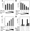
Effect of Rbm15 on Notch-induced HES1 promoter activity is cell type dependent. (A) CHO cells were cotransfected with CMV-Luc along with differing amounts of the GFP-Rbm15 expression plasmid as indicated by the dark black wedge. A GFP expression plasmid was used to maintain a constant DNA amount as indicated by the gray wedge. Data are presented as means ± standard deviations of luciferase activity from triplicate samples from one representative experiment of three that gave similar data. (B) Hes-1-Luc was used as the reporter plasmid, which is activated by NICD via its binding to RBPJκ on the Hes-1 promoter. As in panel A, different amounts of GFP-Rbm15 or GFP expression plasmid were cotransfected into CHO cells with the Hes-Luc and NICD expression plasmids as indicated. (C) 32DWT18 cells were transfected with Hes-Luc and NICD expression plasmids as indicated plus different amounts of GFP and/or GPP-Rbm15 expression plasmids as indicated. Note that a constant amount of DNA was included in every transfection. (D) Relative luciferase activity in 32DWT18 cells transfected as before with Hes-Luc (all lanes), NICD expression plasmid, and plasmids encoding various fragments of Rbm15. Plasmid names are as indicated in Fig. 6B.
FIG. 6.
FIG. 6.
Rbm15 coimmunoprecipitates with RBPJκ. (A) CHO cells were cotransfected with expression plasmids for either GFP or GFP-Rbm15, as indicated, plus expression plasmids for RBPJκ (all lanes). Nuclear extracts were immunoprecipitated with either anti-GFP (lanes 1 and 2) or anti-RBPJκ (lanes 3 and 4) and probed by Western analysis using anti-GFP. Lanes 5 and 6 show unmanipulated input. Bands representing the GFP-Rbm15 fusion protein and GFP are indicated. The band of approximately 60 kDa in lanes 3 and 4 likely represents the anti-RBPJκ antibody. (B) Schematic representation of Rbm15 truncations tested. The full-length (FL) mouse Rbm15 protein contains 962 aa. RNA recognition motifs (RRM) are located at aa 178 to 247, 374 to 446, and 455 to 524. The conserved SPOC domain is located at the C terminus from aa 778 to 957. Nuclear localization signal (NLS) sequences are predicted as shown. The pcDNA N/Rbm15 plasmid contains the 608-aa N-terminal fragment in frame with an HA tag. C/Rbm15 contains a 327-aa C-terminal fragment in frame with a myc tag. Rbm15-F1 through -F4 represent the truncated fragments from the N terminus with 453, 355, 306, and 198 aa, all fused in frame to a V5 tag. (C) Immunoprecipitation (IP) of Flag-tagged RBPJκ (F-RBPJκ) cotransfected with full-length Rbm15, empty pcDNA3, N/Rbm15, pcDNA-myc-tagged empty vector, and C/Rbm15, as indicated. Lysates were immunoprecipitated with anti-GFP, anti-HA, and anti-myc antibodies as shown, and the Western blot (IB) was probed with anti-Flag antibody. (D) Immunoprecipitation of the truncated Rbm15 fragments with anti-Flag antibody as indicated. The upper panel (right side) shows that the truncated Rbm15 polypeptides are all coimmunoprecipitated with RBPJκ (anti-Flag). The middle panel shows that the anti-Flag antibody precipitates the Flag-tagged RBPJκ protein. The bottom panel shows that the anti-Flag antibody in the presence of the Flag-tagged expression plasmid alone does not immunoprecipitate the truncated fragments of Rbm15.

References

    1. Ballerini, P., A. Blaise, T. Mercher, B. Pellegrino, C. Perot, J. van den Akker, E. Gatbois, M. Adam, L. Douay, R. Berger, O. Bernard, and J. Landman-Parker. 2003. A novel real-time RT-PCR assay for quantification of OTT-MAL fusion transcript reliable for diagnosis of t(1;22) and minimal residual disease (MRD) detection. Leukemia 17:1193-1196. - PubMed
    1. Baruchel, A., M. T. Daniel, G. Schaison, and R. Berger. 1991. Nonrandom t(1;22)(p12-p13;q13) in acute megakaryocytic malignant proliferation. Cancer Genet. Cytogenet. 54:239-243. - PubMed
    1. Beatus, P., J. Lundkvist, C. Oberg, and U. Lendahl. 1999. The notch 3 intracellular domain represses notch 1-mediated activation through Hairy/Enhancer of split (HES) promoters. Development 126:3925-3935. - PubMed
    1. Bigas, A., D. I. Martin, and L. A. Milner. 1998. Notch1 and Notch2 inhibit myeloid differentiation in response to different cytokines. Mol. Cell. Biol. 18:2324-2333. - PMC - PubMed
    1. Carroll, A., C. Civin, N. Schneider, G. Dahl, A. Pappo, P. Bowman, A. Emami, S. Gross, C. Alvarado, C. Phillips, et al. 1991. The t(1;22) (p13;q13) is nonrandom and restricted to infants with acute megakaryoblastic leukemia. Blood 78:748-752. - PubMed

Publication types

MeSH terms