. 2007 Apr;27(8):2830-40.
doi: 10.1128/MCB.00079-07. Epub 2007 Feb 5.

The p85alpha regulatory subunit of phosphoinositide 3-kinase potentiates c-Jun N-terminal kinase-mediated insulin resistance

Affiliations

The p85alpha regulatory subunit of phosphoinositide 3-kinase potentiates c-Jun N-terminal kinase-mediated insulin resistance

Cullen M Taniguchi et al. Mol Cell Biol. 2007 Apr.

Abstract

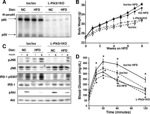
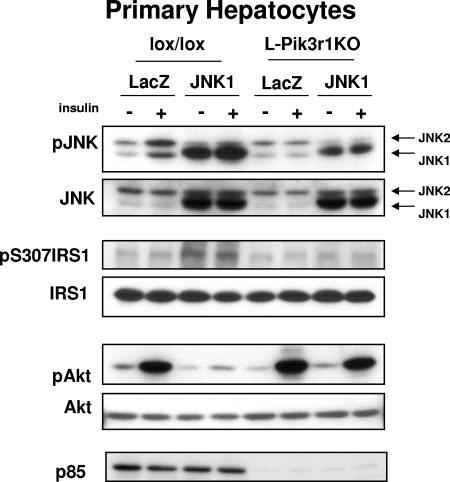
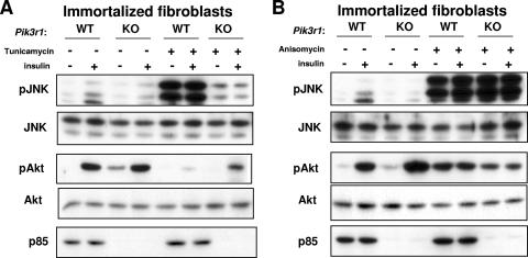
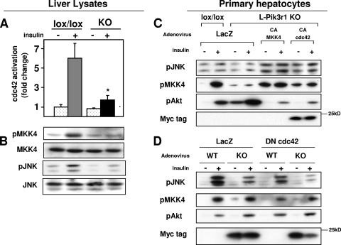
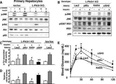
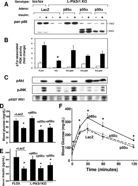
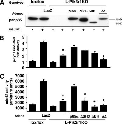
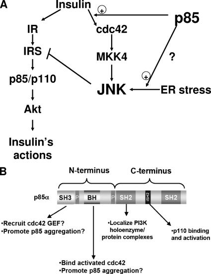
Insulin resistance is a defining feature of type 2 diabetes and the metabolic syndrome. While the molecular mechanisms of insulin resistance are multiple, recent evidence suggests that attenuation of insulin signaling by c-Jun N-terminal kinase (JNK) may be a central part of the pathobiology of insulin resistance. Here we demonstrate that the p85alpha regulatory subunit of phosphoinositide 3-kinase (PI3K), a key mediator of insulin's metabolic actions, is also required for the activation of JNK in states of insulin resistance, including high-fat diet-induced obesity and JNK1 overexpression. The requirement of the p85alpha regulatory subunit for JNK occurs independently of its role as a component of the PI3K heterodimer and occurs only in response to specific stimuli, namely, insulin and tunicamycin, a chemical that induces endoplasmic reticulum stress. We further show that insulin and p85 activate JNK by via cdc42 and MKK4. The activation of this cdc42/JNK pathway requires both an intact N terminus and functional SH2 domains within the C terminus of the p85alpha regulatory subunit. Thus, p85alpha plays a dual role in regulating insulin sensitivity and may mediate cross talk between the PI3K and stress kinase pathways.


Figures

FIG. 1.
FIG. 1.
Loss of hepatic p85α protects mice from obesity-induced diabetes. (A) Western blot assays for expression of p85α and p50α with a pan-p85 antibody in the indicated animals and treatments. IB, immunoblot. (B) Six-week-old L-Pik3r1KO mice and lox/lox controls were fed normal chow (NC) or an HFD for a total of 8 weeks. The graph indicates body weight for each week on either diet. Symbols: open squares, lox/lox, NC; open circles, L-Pik3r1KO, NC; closed squares, lox/lox, HFD; closed circles, L-Pik3r1KO, HFD. (C) Western blot assays were performed against liver lysates of mice with the indicated genotypes and diets with the phosphoserine 473 Akt (pAkt) antibody, phospho-JNK (pJNK) antibody, and phosphoserine 307 (ps307) IRS-1 antibody. The phosphospecific antibody blots were stripped and reprobed with the appropriate antibodies to determine the total levels of the corresponding proteins. (D) GTTs of lox/lox or L-Pik3r1KO mice on NC or an HFD. *, P < 0.05 compared with lox/lox on NC. The error bars represent the SEM (n = 4 to 6).
FIG. 2.
FIG. 2.
L-Pik3r1KO hepatocytes are JNK resistant. Primary hepatocytes were isolated from lox/lox mice or L-Pik3r1KO mice, infected with a JNK1-expressing adenovirus or a control LacZ adenovirus at an MOI of 100, and then treated with saline or 100 nM insulin after 12 h of serum starvation. Western blot assays were then performed against lysates from the treated hepatocytes with the indicated antibodies. The phosphospecific antibody blots were stripped and reprobed with the appropriate antibodies to determine the total levels of the corresponding proteins.
FIG. 3.
FIG. 3.
Cells lacking p85 are resistant to ER stress. (A) WT or Pik3r1/ immortalized fibroblasts were serum starved overnight before a 4-h incubation with tunicamycin as indicated. The cells were then treated with insulin for 15 min as indicated. Lysates from the treated cells were then analyzed by Western blot assay with phospho-JNK and phospho-Akt antibodies. (B) Cells were treated as in panel A, except with a 4-h incubation with anisomycin, as indicated.
FIG. 4.
FIG. 4.
Pik3r1 is required for insulin activation of the cdc42/MKK4/JNK pathway. (A) cdc42 activity as determined by PAK1 pull-down assay from liver lysates 3 min after administration of a 5-U portal bolus of insulin (see Materials and Methods). (B) Phosphospecific-antibody blots against phospho-MKK4 and phospho-JNK. The phosphospecific antibody blots were stripped and reprobed with the appropriate antibodies to determine the total levels of the corresponding proteins. (C) Primary hepatocytes isolated from lox/lox or L-Pik3r1KO mice were infected with either LacZ or constitutively active forms of MKK4 and cdc42 (myc tagged), serum starved overnight, and then stimulated with 100 nM insulin. Western blot assays were then performed with the indicated antibodies. (D) Primary hepatocytes of the indicated genotype were infected with either LacZ or a myc-tagged dominant negative cdc42 adenovirus, serum starved overnight, and then stimulated with 100 nM insulin.
FIG. 5.
FIG. 5.
Activation of cdc42/JNK by p85α is not dependent upon PI3K activity. (A) Primary hepatocytes were isolated from lox/lox or L-Pik3r1KO mice (see Materials and Methods) and treated with insulin or the PI3K inhibitor LY294002 as indicated. JNK and Akt activities were then estimated with phosphospecific antibodies. The phosphospecific antibody blots were stripped and reprobed with the appropriate antibodies to determine the total levels of the corresponding proteins. (B) Purified recombinant adenoviruses (Adeno) were injected via tail vein into 10- to 12-week-old L-Pik3r1KO mice. Mice were injected with adenoviruses encoding control LacZ or WT p85α, RARA p85α, or ΔiSH2 p85α. Western blot assays were then performed against liver lysates of mice treated with adenovirus, with phospho-JNK, phosphoserine307, and pan-p85 antibodies to verify expression levels. Primary hepatocytes were isolated from lox/lox or L-Pik3r1KO mice and infected with the same adenoviruses, i.e., LacZ, WT p85α, RARA p85α, or ΔiSH2 p85α, and then PI3K (C) and cdc42 (D) activities were measured. *, P < 0.05 compared to insulin-stimulated lox/lox (E) GTT of mice whose livers were reconstituted with one of the indicated adenoviruses. The bars represent the SEM (n = 6 to 8).
FIG. 6.
FIG. 6.
The suppression of insulin action by Pik3r1 is specific to the p85α isoform. (A) Recombinant adenoviruses (Adeno) were injected via tail vein into 10- to 12-week-old male mice of the indicated genotypes. Mice were injected with adenoviruses encoding control LacZ or one of the Pik3r1 gene products, p85α, p55α, or p50α. Immunoblot assays were then performed to detect the expression of the p85α isoforms. An extra band of approximately 50 kDa appears in the livers treated with p55α adenovirus, and this likely represents a proteolytic breakdown product of p55α. (B) PI3K activity from mice injected with the indicated adenoviruses. (C) Western blot assays were then performed against liver lysates of mice treated with adenovirus, with phospho-JNK and phosphoserine 307 antibodies. The phosphospecific-antibody blots were stripped and reprobed with the appropriate antibodies to determine the total levels of the corresponding proteins (data not shown). Fasting blood glucose (D) and fasting serum insulin (E) levels of mice treated with the indicated adenoviruses are shown. (F) GTT of mice whose livers were reconstituted with one of the Pik3r1 gene products. The bars represent the SEM (n = 6 to 8).
FIG. 7.
FIG. 7.
The N terminus of p85α is required for cdc42 activation. Primary hepatocytes were isolated from lox/lox or L-Pik3r1KO mice and then infected with control LacZ, WT p85α, ΔSH3, ΔΒΗ, or ΔΔp85α adenovirus (Adeno). (A) Western blot assay with the pan-p85 antibody confirming expression levels of the construct. PI3K activity (B) and cdc42 activity (C) were measured in insulin-stimulated lysates of the infected primary hepatocytes. The bars represent the SEM (n = 3 to 4).
FIG. 8.
FIG. 8.
Molecular mechanisms of JNK activation by p85. (A) Schematic depiction of the role that p85 plays in activating JNK. The full-length p85 regulatory subunit potentiates the activation of JNK through the insulin/cdc42/MKK4 pathway and also facilitates the activation of JNK via the ER stress pathway. Activation of JNK through either pathway results in negative feedback on the insulin signaling pathway. Thus, the loss of p85 improves insulin sensitivity, in part by diminishing this JNK-mediated negative regulation. (B) Schematic of the structural features of the p85 regulatory subunit of PI3K showing how they may impact insulin action. The N terminus contains an SH3 domain, which may mediate intermolecular interactions with possible cdc42 guanine nucleotide exchange factors, while the BH domain binds activated cdc42. The SH2 domains in the C terminus are critical for proper function and localization of PI3K and may also mediate the recruitment of p85α to various cellular complexes. The proline-rich domains (P) are diagrammed, but we have not investigated the roles of these domains in p85α function.

Similar articles

Cited by

References

    1. Antonetti, D. A., P. Algenstaedt, and C. R. Kahn. 1996. Insulin receptor substrate 1 binds two novel splice variants of the regulatory subunit of phosphatidylinositol 3-kinase in muscle and brain. Mol. Cell. Biol. 16:2195-2203. - PMC - PubMed
    1. Backer, J. M., M. G. Myers, Jr., S. E. Shoelson, D. J. Chin, X. J. Sun, M. Miralpeix, P. Hu, B. Margolis, E. Y. Skolnik, J. Schlessinger, and M. F. White. 1992. Phosphatidylinositol 3′-kinase is activated by association with IRS-1 during insulin stimulation. EMBO J. 11:3469-3479. - PMC - PubMed
    1. Bandyopadhyay, G. K., J. G. Yu, J. Ofrecio, and J. M. Olefsky. 2005. Increased p85/55/50 expression and decreased phosphatidylinositol 3-kinase activity in insulin-resistant human skeletal muscle. Diabetes 54:2351-2359. - PubMed
    1. Baud, V., Z. G. Liu, B. Bennett, N. Suzuki, Y. Xia, and M. Karin. 1999. Signaling by proinflammatory cytokines: oligomerization of TRAF2 and TRAF6 is sufficient for JNK and IKK activation and target gene induction via an amino-terminal effector domain. Genes Dev. 13:1297-1308. - PMC - PubMed
    1. Beeton, C. A., P. Das, M. D. Waterfield, and P. R. Shepherd. 1999. The SH3 and BH domains of the p85α adapter subunit play a critical role in regulating class Ia phosphoinositide 3-kinase function. Mol. Cell. Biol. Res. Commun. 1:153-157. - PubMed

Publication types

MeSH terms