. 2007 Apr;27(8):2967-79.
doi: 10.1128/MCB.01830-06. Epub 2007 Feb 5.

Cdh1-anaphase-promoting complex targets Skp2 for destruction in transforming growth factor beta-induced growth inhibition

Affiliations

Cdh1-anaphase-promoting complex targets Skp2 for destruction in transforming growth factor beta-induced growth inhibition

Weijun Liu et al. Mol Cell Biol. 2007 Apr.

Abstract

As a subunit of a ubiquitin ligase, Skp2 is implicated in facilitating cell cycle progression via degradation of various protein targets. We report here that Skp2 is rapidly degraded following cellular stimulation by the cytokine transforming growth factor beta (TGF-beta) and that this degradation stabilizes the cell cycle arrest protein p27. The Skp2 degradation is mediated by Cdh1-anaphase-promoting complex (APC), as shown by depletion of Cdh1 with small interfering RNA, and by reconstitution of ubiquitylation reactions in a purified system. Blockage of Skp2 degradation greatly reduces TGF-beta-induced cell cycle arrest, as does expression of a nondegradable Skp2 mutant. Furthermore, we demonstrate that TGF-beta-induced Skp2 degradation is mediated by the Smad cascade. The degradation of Skp2 stabilizes p27, thereby ensuring TGF-beta-induced cell cycle arrest. These results identify a novel mechanism for tumor suppression by TGF-beta and explain why dysfunction of APC in the TGF-beta pathway in responsive cells is associated with cancer.


Figures
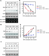
FIG. 1.
FIG. 1.
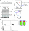
Skp2 is rapidly degraded in response to TGF-β signaling. (A) Skp2 protein levels drop drastically in response to TGF-β stimulation. The half-life of Skp2 in response to TGF-β signaling is about 60 min while the half-life for SnoN is about 30 min. p27 protein levels gradually increase while Skp2 protein levels decrease. Mink lung epithelia cells (Mv1Lu) were treated with TGF-β (100 pM). Protein levels were measured by immunoblotting. Equal amounts of total protein were subjected to immunoblot analysis, as evidenced by the equal concentration of tubulin. Skp2 and actin (control) mRNA levels were monitored by RT-PCR analysis. (B) Skp2 protein chase analysis in response to TGF-β stimulation. Mv1Lu cells were treated with 20 μM cycloheximide. Skp2 protein turnover was measured by immunoblotting. (C) Skp2 degradation is blocked by the proteasomal inhibitor MG-132 (50 μM). (D) Quantification of Skp2 protein levels in response to TGF-β in the presence or absence of cycloheximide or MG-132. (E) Quantification of p27 protein levels in response to TGF-β in the presence or absence of cycloheximide or MG-132. CHX, cycloheximide.
FIG. 2.
FIG. 2.
Cdh1 is necessary and sufficient for downregulation of Skp2 in response to TGF-β stimulation. (A) Creation of Cdh1 siRNA stable clones. Based on Cdh1 knock-down results using transient transfection of siRNA duplex, two oligonucleotides (N terminus, residues 338 to 358; C terminus, residues 566 to 586) were chosen for construction of Cdh1 siRNA stable cells. Cell extracts (100 μg) were blotted with Cdh1 and PCNA (control) antibodies. Cdh1 levels in clone 3 (using an N-terminal oligonucleotide) and clone 6 (using a C-terminal oligonucleotide) were reduced approximately 90%, while Cdh1 levels did not fall in clone 1 (a control clone generated using firefly luciferase siRNA). (B) Depletion of Cdh1 blocks Skp2 degradation in response to TGF-β stimulation. (C) Quantification of Skp2 and p27 protein levels in response to TGF-β signaling in wild-type and Cdh1 siRNA cells. (D) Knock-down of Cdh1 antagonizes TGF-β-induced G1 arrest. Wild-type and Cdh1 siRNA cells were treated with TGF-β. The cell cycle profile was measured by fluorescence-activated cell sorting analysis 20 h after the stimulation with TGF-β. (E) Depletion of Cdh1 blocks the TGF-β-induced cell growth inhibition. Wild-type and Cdh1 siRNA cells were treated with TGF-β at different concentrations. Cell numbers were counted 72 h after the stimulation with TGF-β. WT, wild type.
FIG. 3.
FIG. 3.
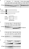
Degradation of Skp2 induced by TGF-β is mediated by Cdh1-APC. (A) Skp2 degradation is induced by TGF-β stimulation. Mv1Lu cells were treated (or not treated) with 100 pM TGF-β for 60 min, and extracts were prepared. 35S-labeled in vitro translated Skp2 was added to the extracts and supplemented with protein degradation cocktail. Aliquots were removed at the indicated times and resolved by SDS-PAGE. Skp2 degradation was measured by autoradiography. (B) Alignment of Skp2 with known APC substrates, cyclin B, and securin. Similar to other substrates of APC, Skp2 contains a conserved destruction box [RXXLXXX(N/D)] at its NH2 terminus. (C) Destruction box is required to mediate TGF-β-induced Skp2 degradation. Wild-type and mutant Skp2 proteins lacking the destruction box were incubated in TGF-β-stimulated extracts. (D) Depletion of Cdh1 blocks Skp2 degradation in TGF-β-stimulated extracts. Skp2 was incubated with the extracts prepared from wild-type Mv1Lu or Cdh1 siRNA cells. (E) Degradation of Skp2 is blocked by the addition of a destruction box-containing peptide. NH2-terminal fragments (amino acids 1 to 102) of Xenopus cyclin B or a control fragment lacking the destruction box [CycB-Δdb (N102)] were added to the TGF-β-stimulated extract and 35S-labeled in vitro translated Skp2 at the indicated concentrations. After incubation for 90 min at room temperature, samples were analyzed by SDS-PAGE and autoradiography.
FIG. 4.
FIG. 4.
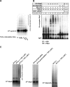
Skp2 is ubiquitylated in vivo and in a purified system. (A) Activity of APC is enhanced following stimulation by TGF-β. Mv1Lu cells were stimulated with TGF-β and harvested at different times as indicated. APC was subsequently purified from the cell lysate using anti-Cdc27 antibody coupled to protein A beads and was subjected to ubiquitylation mixture for a ubiquitylation assay. 35S-labeled in vitro translated cyclin B was used as a putative substrate for APC activity analysis. Polyubiquitin conjugates of cyclin B were measured by autoradiography. (B) Skp2 is polyubiquitylated in vivo. Myc-tagged ubiquitin and HA-tagged wild-type Skp2 or mutant Skp2 lacking the destruction box were cotransfected into wild-type or Cdh1 siRNA Mv1Lu cells. Cells were treated with TGF-β and harvested at the indicated times. Skp2 complex was immuno-purified by anti-HA antibody. The polyubiquitin-conjugated Skp2 was detected by immunoblotting using anti-Myc antibody. (C) Recapitulation of Skp2 ubiquitylation in a purified system. Mv1Lu cells were stimulated with TGF-β and harvested at different times as indicated. APC was purified from the cell lysate using anti-Cdc27 antibody coupled to protein A beads and was subjected to a ubiquitylation mixture for the ubiquitylation assay. Polyubiquitin conjugates of Skp2 were measured by autoradiography. Depletion of Cdh1 attenuates TGF-β induced Skp2 ubiquitylation. In addition, disruption of the destruction box blocks TGF-β-induced Skp2 ubiquitylation. IP, immunoprecipitation; IB, immunoblotting; α, anti.
FIG. 5.
FIG. 5.
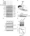
TGF-β-induced Skp2 degradation is mediated by a Smad cascade. (A) Skp2 forms a complex with Cdh1 in response to TGF-β signaling. Mv1Lu cells were cotransfected with expression vectors encoding HA-Skp2, Myc-Cdh1, Flag-Smad3, and V5-Cdc27. Transfected cells were preincubated with the proteasomal inhibitor, MG-132, or vehicle (DMSO) for 1 h before stimulation with TGF-β. HA-Skp2 complexes were recovered using an anti-HA matrix. The interaction among Skp2, Cdh1, Smad3, and Cdc27 was analyzed by protein immunoblotting with anti-HA, anti-Myc, anti-Flag, and anti-V5. Expression levels of HA-Skp2, Myc-Cdh1, Flag-Smad3, and V5-Cdc27 were determined by immunoblotting. (B) Diagram for TGF-β-induced quaternary complex of Skp2, Cdh1, Smad3, and Cdc27. (C) Depletion of Smad3 by transfection of Smad3 siRNA oligonucleotides. (D) Depletion of Smad3 protein attenuated TGF-β-induced Skp2 degradation. (E) Knock-down of Smad3 blocked TGF-β-induced Skp2 ubiquitylation. (F) Depletion of Smad3 antagonized TGF-β-induced growth inhibition. IP, immunoprecipitation.
FIG. 6.
FIG. 6.
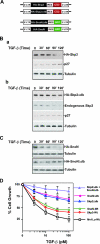
Expression of nondegradable Skp2 and SnoN antagonizes TGF-β signaling. (A) Diagram of retroviral vector expressing nondegradable Skp2 and SnoN. (B) Protein levels of stably expressed wild-type Skp2 (a) and nondegradable Skp2 (b) in response to TGF-β signaling. (C) Protein levels of stably expressed wild-type SnoN and nondegradable SnoN in response to TGF-β signaling. (D) Growth inhibition assay. Mv1Lu cells stably expressing Skp2, Skp2Δdb, SnoN, and SnoNΔdb, respectively, and coexpressing Skp2Δdb and SnoNΔdb were incubated for 4 days with various concentrations of TGF-β1 as indicated. The growth of cells was quantified by cell counting and compared with the growth of unstimulated cells. WT, wild type.
FIG. 7.
FIG. 7.
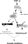
Model for the role of Cdh1-APC in TGF-β-induced degradation of Skp2 and SnoN. APC is activated by TGF-β stimulation. Activated APC then targets Skp2 for degradation, thereby stabilizing p27. In addition, activated APC is required for SnoN degradation, hence permitting transactivation of TGF-β-responsive tumor suppressor genes. Thus, both induction of tumor suppressors and stabilization of p27 are necessary to achieve cell cycle arrest.

Similar articles

Cited by

References

    1. Ang, X. L., and J. W. Harper. 2004. Interwoven ubiquitination oscillators and control of cell cycle transitions. Sci. STKE 2004:e31. - PubMed
    1. Attisano, L., J. L. Wrana, E. Montalvo, and J. Massague. 1996. Activation of signalling by the activin receptor complex. Mol. Cell. Biol. 16:1066-1073. - PMC - PubMed
    1. Bashir, T., N. V. Dorrello, V. Amador, D. Guardavaccaro, and M. Pagano. 2004. Control of the SCF (Skp2-Cks1) ubiquitin ligase by the APC/C (Cdh1) ubiquitin ligase. Nature 428:190-193. - PubMed
    1. Carrano, A. C., E. Eytan, A. Hershko, and M. Pagano. 1999. SKP2 is required for ubiquitin-mediated degradation of the CDK inhibitor p27. Nat. Cell Biol. 1:193-199. - PubMed
    1. Catzavelos, C., N. Bhattacharya, Y. C. Ung, J. A. Wilson, L. Roncari, C. Sandhu, P. Shaw, H. Yeger, I. Morava-Protzner, L. Kapusta, E. Franssen, K. I. Pritchard, and J. M. Slingerland. 1997. Decreased levels of the cell-cycle inhibitor p27Kip1 protein: prognostic implications in primary breast cancer. Nat. Med. 3:227-230. - PubMed

Publication types

MeSH terms

Substances