. 2007 Apr 6;282(14):10576-84.
doi: 10.1074/jbc.M609695200. Epub 2007 Feb 5.

Use of a cAMP BRET sensor to characterize a novel regulation of cAMP by the sphingosine 1-phosphate/G13 pathway

Affiliations

Use of a cAMP BRET sensor to characterize a novel regulation of cAMP by the sphingosine 1-phosphate/G13 pathway

Lily I Jiang et al. J Biol Chem. .

Abstract

Regulation of intracellular cyclic adenosine 3 ',5 '-monophosphate (cAMP) is integral in mediating cell growth, cell differentiation, and immune responses in hematopoietic cells. To facilitate studies of cAMP regulation we developed a BRET (bioluminescence resonance energy transfer) sensor for cAMP, CAMYEL (cAMP sensor using YFP-Epac-RLuc), which can quantitatively and rapidly monitor intracellular concentrations of cAMP in vivo. This sensor was used to characterize three distinct pathways for modulation of cAMP synthesis stimulated by presumed G(s)-dependent receptors for isoproterenol and prostaglandin E(2). Whereas two ligands, uridine 5 '-diphosphate and complement C5a, appear to use known mechanisms for augmentation of cAMP via G(q)/calcium and G(i), the action of sphingosine 1-phosphate (S1P) is novel. In these cells, S1P, a biologically active lysophospholipid, greatly enhances increases in intracellular cAMP triggered by the ligands for G(s)-coupled receptors while having only a minimal effect by itself. The enhancement of cAMP by S1P is resistant to pertussis toxin and independent of intracellular calcium. Studies with RNAi and chemical perturbations demonstrate that the effect of S1P is mediated by the S1P(2) receptor and the heterotrimeric G(13) protein. Thus in these macrophage cells, all four major classes of G proteins can regulate intracellular cAMP.


Figures

FIGURE 1
FIGURE 1. An Epac-based BRET sensor for cAMP (CAMYEL)
A, schematic drawing of the domain structure of CAMYEL. The inactive cytosolic mutant form of human Epac-1 (amino acids 149−881, T781A, F782A) (21) was flanked by citrine or a circularly permuted citrine-cp229 and Renilla luciferase (RL). B and C, comparison of emission spectra of Citrine-Epac-RL (B) and Citrine-cp229-Epac-RL (C) in the presence and absence of 100 μm cAMP. HEK293 cells were transiently transfected with either construct. Cells were lysed with buffer containing 20 mm NaHEPES, pH 7.4, 50 mm KCl, 50 mm NaCl, 2.5 mm MgCl2, 0.2% Nonidet P-40, 5 mm dithiothreitol, and protease inhibitors. Emission spectra were measured in the presence of 2 μm coelenterazine-h substrate with or without 100 μm cAMP.
FIGURE 2
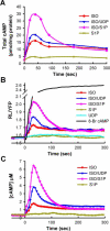
FIGURE 2. Measurement of intracellular cAMP responses in RAW 264.7 cells
A, changes in cAMP measured by EIA. At time 0, cells were stimulated with 16 nm ISO, 16 nm ISO and 0.5 μm UDP, 16 nm ISO and 10 nm S1P, or 50 nm S1P as indicated. At the given times, reactions were stopped by removal of medium and addition of cell lysis solution, and cAMP was determined by EIA. The results shown are the average of three independent experiments done with two different preparations of cells: the variance among the experiments is about 30% of the signals. B, measurement of cAMP responses using the CAMYEL sensor. Emission ratios (RL/YFP) were measured in RAW 264.7 cells stably expressing the CAMYEL sensor. Cells were stimulated at time 0 by addition of either 16 nm ISO 16 nm ISO and 0.5 μm UDP, 16 nm ISO and 10 nm S1P, 10 nm S1P, 2.5 μm UDP, or 2 mm 8-bromo cAMP as indicated. C, ratiometric measurements of CAMYEL responses were converted to intracellular concentrations of cAMP.
FIGURE 3
FIGURE 3. Effect of S1P on intracellular cAMP has a fast onset and is not affected by inhibitors of phosphodiesterases
The CAMYEL sensor was used to measure the rise in cAMP when cells were treated with 16 nm ISO or 16 nm ISO and 10 nm S1P either in the absence or in the presence of the phosphodiesterase inhibitors (PDEi), 10 μm Ro20−1724 and 40 μm isobutylmethylxanthine. Ligands were added at time 0, PDEi were added 2 min prior to ligand addition, which was sufficient to cause maximum impact of the inhibitors.
FIGURE 4
FIGURE 4. Effect of S1P on cAMP parallels activation of Gs
A, changes in cAMP were measured with CAMYEL; cells were treated with 16 nm ISO at time 0 followed by addition of 10 nm S1P at the indicated times. B, effect of S1P on intracellular cAMP is expressed as the ratio of the S1P induced peak of cAMP to the concentration of cAMP induced by ISO alone at the time of S1P addition. For simultaneous addition of ISO and S1P the ratio is calculated as the peak induced by the two ligands versus that by ISO alone. The synergistic effect of S1P on cAMP appears to be the same throughout the 7 min assay period. C, termination of β-adrenergic receptor signaling diminished the effect of S1P. All cells were treated with 2.5 μm TER at time 0. Addition of 10 nm S1P at 30 s greatly enhanced the cAMP response stimulated by TER (cyan, x). Simultaneous addition of 10 nm S1P and 10 μm ICI 118551 (ICI), a β-adrenergic antagonist, greatly reduced the ability of S1P to affect increases in cAMP (red, ○). Addition of 10 μm ICI 118551 at 30 s immediately initiated a decline in cAMP; subsequent addition of 10 nm S1P 12 s later produced no effect on intracellular cAMP concentration (green, ▽).
FIGURE 5
FIGURE 5. Effect of intracellular calcium and activity of the Gi pathway on cAMP responses to dual ligands in RAW 264.7 cells
Cells expressing the CAMYEL sensor were treated with 50 ng/ml pertussis toxin for 20 h or 1 μm thapsigargin and 2 mm EGTA for 2 min as indicated prior to ligand additions. cAMP responses were induced by addition of 16 nm ISO at time 0 followed by addition of 10 nm S1P (A), 0.5 μm UDP (B), or 100 nm C5a (C). Results shown are averages from at least three experiments, error bars represent the S.D.
FIGURE 6
FIGURE 6. The S1P2 receptor transduces signaling by S1P for modulation of intracellular cAMP
A, RAW cells expressing CAMYEL were transiently transfected with control siRNA or pools of four siRNA oligomers (designated as –P) targeting either the S1P1 or S1P2 receptor. Analysis by qRT-PCR showed specific but partial knockdowns of the S1P1 and S1P2 receptors. B–D, cells were assayed for cAMP responses to simultaneous addition of 16 nm ISO and 0.5 μM UDP (B), simultaneous addition of 16 nm ISO and 10 nm S1P (C), and sequential addition of 16 nm ISO followed by 10 nm S1P (D). Knockdown of S1P2 using a single oligomer (S1P2-B) that is independent of the pool of four oligomers resulted in a similar phenotype (D). E, addition of 1 μm SEW2871, an agonist for the S1P1 receptor, at 2 min after addition of ISO failed to induce a synergistic cAMP response. F, addition of 100 nm JTE-013, an antagonist of S1P2 receptors, at 12 s prior to addition of S1P significantly reduced the synergistic effect on cAMP. Ligands were added at time 0 or as indicated. Error bars represent the S.D. of results from four experiments of 2 independent transfections. Errors were similar for the other traces but left out for clarity.
FIGURE 7
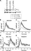
FIGURE 7. Gα13 is required for the synergy of S1P on Gs-mediated cAMP responses
A, RAW cells expressing CAMYEL were transiently transfected with control siRNA or a pool of four siRNA oligomers (designated as –P) specifically targeting Gα13. The presence of Gα13 was assessed by Western blot. The reduction of the α-subunit in cells treated with four siRNA oligomers targeting Gα13 was about 60%. B–D, response of cAMP in control transfected and Gα13 knockdown cells to simultaneous addition of 16 nm ISO and 0.5 μm UDP (B), simultaneous addition of 16 nm ISO and 10 nm S1P (C), and sequential addition of 16 nm ISO followed by 10 nm S1P (D). E, synergy of S1P on intracellular cAMP induced by PGE is also reduced in Gα13 knockdown cells. Ligands were added at time 0 or as indicated. Error bars represent the standard deviation of results from four experiments of 2 independent transfections. Errors were similar for the other traces but left out for clarity.

Similar articles

Cited by

References

    1. Lania A, Mantovani G, Spada A. Eur. J. Endocrinol. 2001;145:543–559. - PubMed
    1. Castro A, Jerez MJ, Gil C, Martinez A. Med. Res. Rev. 2005;25:229–244. - PubMed
    1. Lerner A, Epstein P. Biochem. J. 2006;393:21–41. - PMC - PubMed
    1. Wang H, Storm DR. Mol. Pharmacol. 2003;63:463–468. - PubMed
    1. Movsesian MA, Bristow MR. Curr. Top. Dev. Biol. 2005;68:25–48. - PubMed

Publication types

MeSH terms