. 2007 Apr;75(4):1745-50.
doi: 10.1128/IAI.01528-06. Epub 2007 Feb 5.

Evidence for widespread epithelial damage and coincident production of monocyte chemotactic protein 1 in a murine model of intestinal ricin intoxication

Affiliations

Evidence for widespread epithelial damage and coincident production of monocyte chemotactic protein 1 in a murine model of intestinal ricin intoxication

J Marina Yoder et al. Infect Immun. 2007 Apr.

Abstract

The development of small-animal models is necessary to understand host responses and immunity to emerging infectious diseases and potential bioterrorism agents. In this report we have characterized a murine model of intestinal ricin intoxication. Ricin administered intragastrically (i.g.) to BALB/c mice at doses ranging from 1 to 10 mg/kg of body weight induced dose-dependent morphological changes in the proximal small intestine (i.e., duodenum), including widespread villus atrophy and epithelial damage. Coincident with epithelial damage was a localized increase in monocyte chemotactic protein 1, a chemokine known to be associated with inflammation of the intestinal mucosa. Immunity to intestinal ricin intoxication was achieved by immunizing mice i.g. with ricin toxoid and correlated with elevated levels of antitoxin mucosal immunoglobulin A (IgA) and serum IgG antibodies. We expect that this model will serve as a valuable tool in identifying the inflammatory pathways and protective immune responses that are elicited in the intestinal mucosa following ricin exposure and will prove useful in the evaluation of antitoxin vaccines and therapeutics.


Figures
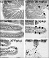
FIG. 1.
FIG. 1.
Histological changes in the murine duodenum associated with ricin intoxication. BALB/c mice were administered PBS (control) (a and c) or ricin (b and d to f) at the indicated doses by gavage and were sacrificed 18 h later. Intestinal segments (1 to 2 cm) were immersed in Bouin's fixative and embedded in paraffin. Tissue sections (5 μm) were stained with H&E and viewed by bright-field microscopy. (a) Low-power (×10) image of a cross section of the duodenum of a control, PBS-fed mouse. The villi appear as long, slender, finger-like projections extending into the intestinal lumen. (b) Low-power image of a cross-section of the duodenum from a mouse treated with ricin (10 mg/kg). The villi appear blunted and swollen, especially at the tips. The swelling (arrowheads) is likely due to edema. (c) High-power (×40) image of two villi from the duodenum of a control, PBS-treated mouse. (d) High-power image of a villus from the duodenum of a mouse treated with ricin (5 mg/kg). There is extensive interepithelial swelling (arrowheads) along the basolateral aspects of enterocytes. Excessive mucus, which appears as “webbing” in the lumen surrounding the affected villus, is also evident. (e) High-power image of two villi from a mouse exposed to ricin (10 mg/kg). At the tips of the villi, the epithelium has separated from the lamina propria (arrowheads), and the basal aspects of many individual enterocytes have degenerated or have been completed destroyed. (f) Cellular infiltrate, consisting of polymorphonuclear leukocytes (arrows), was occasionally evident in the intestinal lumen of ricin-treated animals.
FIG. 2.
FIG. 2.
Histological changes in the murine duodenum associated with ricin intoxication. Paraffin sections of duodena collected from mice 24 h after the animals were challenged i.g. with ricin at the indicated doses (0 to 10 mg/kg) were scored by light microscopy. The tissues were scored based on villus morphology, interepithelial swelling, and cellular infiltrate in the lumen. At least 40 sections from 2 mice were examined at each dose. Average scores with SE are shown.
FIG. 3.
FIG. 3.
Dose- and time-dependent increases in MCP-1 levels in the duodena of mice challenged with ricin. The duodena of BALB/c mice challenged i.g. with ricin were homogenized and assayed for MCP-1 by CBA. (A) Dose-dependent MCP-1 production. Tissues were collected 24 h after groups of mice (n = 5/group) had been challenged with the indicated doses of ricin. Average values with SE are shown. (B) Time-dependent MCP-1 production. Groups of mice (n = 6) were challenged with ricin (5 mg/kg), and tissues were then collected at the indicated time points. Average values with SE are shown. The amount of total protein in each intestinal homogenate sample was determined using the bicinchoninic acid assay (Pierce Chemical). Statistical analysis of differences between groups of mice was determined using an independent t test.
FIG. 4.
FIG. 4.
Antiricin IgG and IgA titers in serum and fecal samples from mice immunized i.g. with RT. BALB/c mice (n = 6 per group) were immunized i.g. with PBS (control) or RT three times (days 0, 14, and 36), and serum and fecal samples were collected 7 days later. Antiricin-specific antibody titers were determined using a ricin-specific ELISA and were expressed as n-fold increases over baseline titers measure in PBS-immunized littermates, as described in Materials and Methods. (A) Antiricin-specific IgG levels in serum. (B) Antiricin-specific IgA levels in serum and fecal extracts.
FIG. 5.
FIG. 5.
Mice immunized i.g. with RT were protected against subsequent ricin challenge. Groups of mice were immunized i.g. five times at approximately 2-week intervals with PBS or RT and then challenged i.g. with ricin (5 mg/kg). Twenty-four hours after challenge, the animals were sacrificed, and the duodena were analyzed for histological changes and MCP-1 levels, as described in the legends to Fig. 2 and 3, respectively. (A) Mice immunized with PBS and challenged with ricin (PBS/ricin) had significantly (P = 0.01) more lesions than control animals (PBS/PBS) or animals immunized with RT and then challenged with ricin (RT/ricin). (B) MCP-1 levels were significantly (P = 0.02) elevated in PBS/ricin animals, compared to RT/ricin and PBS/PBS animals. Statistical significance was determined using an independent t test.

Similar articles

Cited by

References

    1. Anonymous. 2004. Summary of the NIAID Ricin Expert Panel Workshop. National Institutes of Health, Bethesda, MD.
    1. Atlas, R. M. 2003. Bioterrorism and biodefence research: changing the focus of microbiology. Nat. Rev. Microbiol. 1:70-74. - PubMed
    1. Audi, J., M. Belson, M. Patel, J. Schier, and J. Osterloh. 2005. Ricin poisoning: a comprehensive review. JAMA 294:2342-2351. - PubMed
    1. Baenziger, J. U., and D. Fiete. 1979. Structural determinants of Ricinus communis agglutinin and toxin specificity for oligosaccharides. J. Biol. Chem. 254:9795-9799. - PubMed
    1. Bischoff, S. C., M. Krieger, T. Brunner, and C. A. Dahinden. 1992. Monocyte chemotactic protein 1 is a potent activator of human basophils. J. Exp. Med. 175:1271-1275. - PMC - PubMed

Publication types

MeSH terms