. 2007 Feb 12;176(4):483-95.
doi: 10.1083/jcb.200608137. Epub 2007 Feb 5.

Midbody and primary cilium of neural progenitors release extracellular membrane particles enriched in the stem cell marker prominin-1

Affiliations

Midbody and primary cilium of neural progenitors release extracellular membrane particles enriched in the stem cell marker prominin-1

Véronique Dubreuil et al. J Cell Biol. .

Abstract

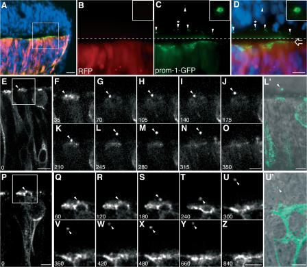
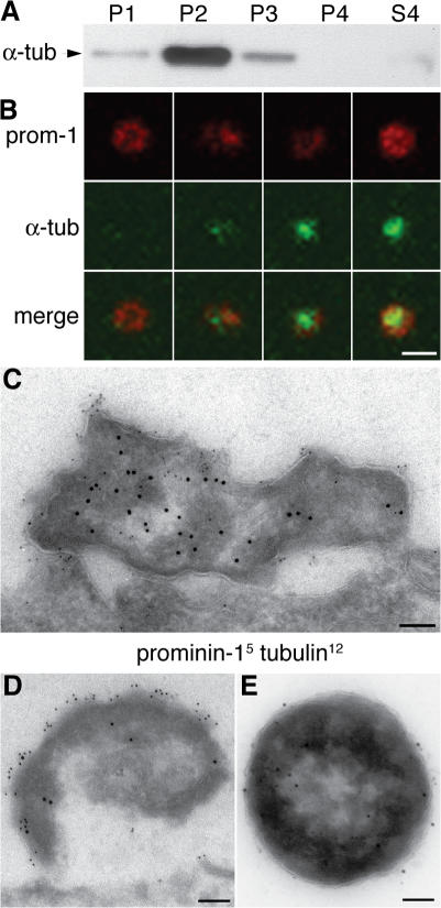
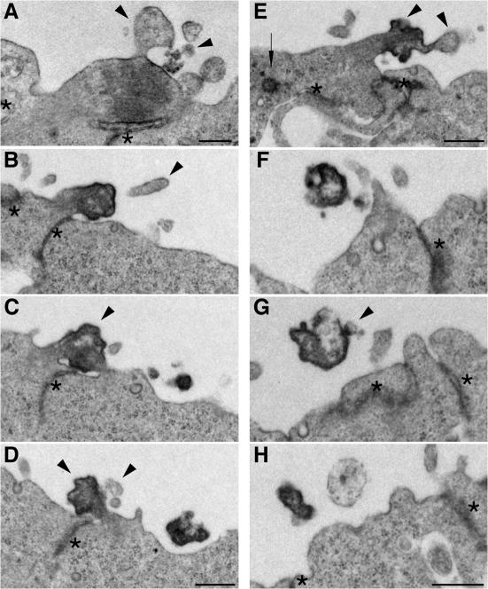
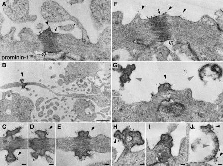
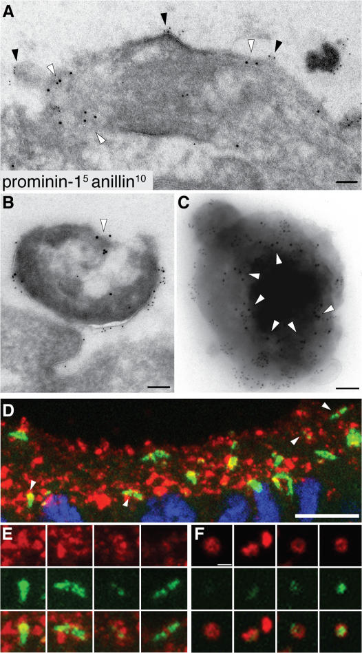
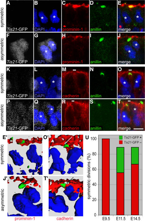
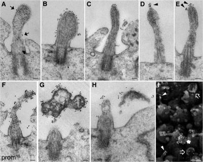
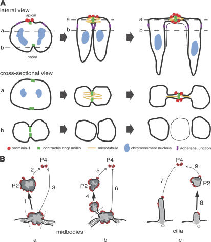
Expansion of the neocortex requires symmetric divisions of neuroepithelial cells, the primary progenitor cells of the developing mammalian central nervous system. Symmetrically dividing neuroepithelial cells are known to form a midbody at their apical (rather than lateral) surface. We show that apical midbodies of neuroepithelial cells concentrate prominin-1 (CD133), a somatic stem cell marker and defining constituent of a specific plasma membrane microdomain. Moreover, these apical midbodies are released, as a whole or in part, into the extracellular space, yielding the prominin-1-enriched membrane particles found in the neural tube fluid. The primary cilium of neuroepithelial cells also concentrates prominin-1 and appears to be a second source of the prominin-1-bearing extracellular membrane particles. Our data reveal novel origins of extracellular membrane traffic that enable neural stem and progenitor cells to avoid the asymmetric inheritance of the midbody observed for other cells and, by releasing a stem cell membrane microdomain, to potentially influence the balance of their proliferation versus differentiation.


Figures

Figure 1.
Figure 1.
Release of prom1-GFP–bearing particles from the apical surface of NE cells. Chick spinal cord (HH10–11) was coelectroporated with prom1-GFP and mRFP and analyzed 24 h later. (A–D) Transverse cryosection of fixed spinal cord analyzed by conventional fluorescence microscopy. DAPI is in blue, mRFP is in red, and prom1-GFP is in green. (A) Low-magnification overview. (B–D) Higher magnification of mRFP (B) and prom1-GFP (C) fluorescence in the area indicated by the white square in A; the merge in D includes the DAPI staining of nuclei. The top half of the images (i.e., above the dashed line) has been contrast enhanced for mRFP and prom1-GFP to facilitate detection of the prom1-GFP–bearing particles at the apical surface of, and within, the nontransfected, contralateral side of the neuroepithelium (arrowheads). The open arrow indicates the apical surface of transfected side of the neuroepithelium. The insets show a higher magnification of the prom1-GFP–bearing particle indicated by the arrowheads with asterisks. (E–Z) Slice cultures were subjected to time-lapse confocal imaging of prom1-GFP, using either 35-s (E–O) or 60-s (P–Z) intervals. The time points shown are indicated in the bottom left corner of each panel (in seconds). Selected single optical sections, following the prom1-GFP–bearing particle (arrowheads) through the z stack, are shown. The apical surface of the transfected side of the neuroepithelium is up. In E, note the basal position of the nucleus of the NE cell releasing the prom1-GFP–bearing particle. (F–O and Q–Z) Higher magnification of the area indicated by the white rectangle in E and P, respectively. (L′ and U′) Overlay of the DIC image and the prom1-GFP fluorescence (green) corresponding to L and U, respectively; note the localization of the prom1-GFP–bearing particle in the lumen of the neural tube (L′) and in the contralateral neuroepithelium (U′). Bars: (A) 20 μm; (D and E) 10 μm; (P, O, Z, L′, and U′) 5 μm.
Figure 2.
Figure 2.
Prom1 particles contain α-tubulin. (A) Neural tube fluid of E11.5 mouse embryos was subjected to differential centrifugation (Marzesco et al., 2005) followed by immunoblot analysis of the fractions for α-tubulin. P1, 300-g pellet; P2, 1,200-g pellet; P3, 10,000-g pellet; P4, 100,000-g pellet; S4, 100,000-g supernatant. (B) Transverse cryosection of E10.5 mouse telencephalon double immunostained for prom1 (red) and α-tubulin (green) and analyzed by confocal microscopy; four examples of prom1 particles in the lumen of the telencephalic ventricle are shown (z-stack projection). (C and D) Transverse ultrathin cryosections of the apical region of mouse E10.5 telencephalic NE cells immunogold labeled for prom1 (5 nm) and α-tubulin (12 nm) showing two doubly immunoreactive particles at the lumenal surface. (E) Negative staining of a particle from the P2 pellet after immunogold labeling for prom1 (5 nm) and acetylated tubulin (12 nm). Bars: (B) 1 μm; (C–E) 100 nm.
Figure 3.
Figure 3.
Ultrastructural resemblance of lumenal particles to aged midbodies of NE cells. EM analysis of serial ultrathin (70 nm) plastic sections of the apical surface of E10.5 mouse telencephalic neuroepithelium. (A) Midbody connecting NE daughter cells in telophase. (B–D) Series showing every other section of an aged midbody. (E) Midbody of an NE cell that has relocated the centriole (arrow) apically. (F–H) Series showing every third section of an electron-dense particle detached from the apical surface of the neuroepithelium. Arrowheads indicate plasma membrane buds and protrusions, and asterisks indicate adherens junctions. The complete sequence of serial sections from which A, E, and F–H were selected is shown in Fig. S4 (A–C), respectively (available at http://www.jcb.org/cgi/content/full/jcb.200608137/DC1). Bars, 500 nm.
Figure 4.
Figure 4.
Prom1 is concentrated at the midbody of NE cells. Mouse embryonic forebrain was subjected to preembedding immunogold labeling for prom1 (10 nm gold) followed by EM analysis of ultrathin serial plastic sections. Apical midbodies of NE cells at E8.5 (A–E), E10.5 (H–J), and E12.5 (F and G) are shown. (B and C) The central portion of the midbody with long, thin stalks depicted in B is shown at higher magnification of a consecutive section in C. (D and E) Consecutive sections showing the central portion of another midbody with long, thin stalks. (G–I) Midbodies with short stalks. (J) Particle at the apical surface of a neuroephithelial cell. Black arrowheads indicate prom1-labeled plasma membrane buds and protrusions, gray arrowheads indicate detached particles, solid arrows indicate midbody plasma membrane facing the lumenal side, and open arrows indicate midbody plasma membrane derived from cleavage furrow. Bars: (A and C–J) 100 nm; (B) 1 μm.
Figure 5.
Figure 5.
Anillin is present not only in prom1-bearing midbodies but also in prom1-bearing lumenal particles. (A and B) Transverse ultrathin cryosections of the apical surface of E10.5 mouse telencephalic neuroepithelium double immunogold labeled for prom1 (5 nm) and anillin (10 nm; white arrowheads). Doubly immunoreactive midbody (A) and particle at the apical surface of the neuroepithelium (B). Black arrowheads indicate prom1-labeled regions at the lumenal plasma membrane of the midbody. (C) Double immunogold labeling for prom1 (5 nm) and anillin (10 nm; white arrowheads) of a particle from the P2 pellet of E11.5 neural tube fluid. (D–F) Transverse cryosections of E11.5 (D and E) and E10.5 (F) mouse telencephalon double immunostained for prom1 (red) and anillin (green) and analyzed by confocal microscopy. (D and E) Z-stack projection providing an en face view onto the apical surface of the neuroepithelium; for orientation, the DAPI staining of nuclei (blue) is shown for one of the optical sections in D. (E) Selected regions of D (indicated by arrowheads) at higher magnification. (F) Four examples of prom1-bearing particles in the lumen of the telencephalic ventricle (z-stack projection). Bars: (A–C) 100 nm; (D) 10 μm; (F) 1 μm.
Figure 6.
Figure 6.
Localization of anillin relative to prom1 and cadherin in Tis21-GFP–negative versus –positive mitotic NE cells. (A–T) Transverse cryosections of E11.5 forebrain of Tis21-GFP knockin mice (GFP expression; white) double immunostained for anillin (green) and either prom1 (A–J; red) or cadherin (K–T; red) and analyzed by confocal microscopy (C–E, H–J, M–O, and R–T, single optical sections; A, B, F, G, K, L, P, and Q, projection of four consecutive optical sections). Nuclei are stained by DAPI (blue). White asterisks indicate telophase nuclei of daughter cells. Small white bars in M, O, R, and T indicate cadherin hole. (A–E and K–O) Telophase NE cells lacking Tis21-GFP expression and undergoing symmetric division (anillin colocalized with prom1 and cadherin hole upon 90–100% complete ingression of the cleavage furrow). (F–J and P–T) Telophase NE cells showing strong (F) or weak (P) Tis21-GFP expression and undergoing asymmetric division (anillin distinct from prom1 and colocalized with cadherin upon 90–100% complete ingression of the cleavage furrow). (E′, J′, O′, and T′) 3D reconstruction from six to eight 1-μm optical sections showing the prom1–anillin–DAPI merge of the symmetrically dividing Tis21-GFP–negative cell in E (E′), the asymmetrically dividing Tis21-GFP–positive cell in J (J′), the cadherin–anillin–DAPI merge of the symmetrically dividing Tis21-GFP–negative cell in O (O′), and the asymmetrically dividing Tis21-GFP–positive cell in T (T′). White and black arrows in O′ and T′, respectively, indicate the cadherin hole. (U) Quantitation of anillin-stained apical midbodies in symmetrically versus asymmetrically dividing NE cells. Forebrain NE cells of E9.5, E11.5, and E14.5 Tis21-GFP knockin mice were double immunostained for cadherin and anillin and analyzed by confocal microscopy as in K–T. Telophase NE cells showing 90–100% complete ingression of the cleavage furrow (25 out of 54 cells analyzed) were first scored for colocalization of the apical anillin staining with either cadherin-negative (symmetric division) or cadherin-positive (asymmetric division) segments of the cell surface and then for the absence or presence of Tis21-GFP expression. Symmetrically dividing NE cells are expressed as a percentage of symmetrically dividing plus asymmetrically dividing cells; the percentage of symmetrically dividing Tis21-GFP–positive cells is indicated in green. Bar, 5 μm.
Figure 7.
Figure 7.
Prom1 is concentrated on cilia of NE cells. Mouse forebrain was subjected to preembedding immunogold labeling for prom1 (10 nm gold) followed by EM analysis of ultrathin plastic sections. (A) A cilium at E8.5 showing weak labeling (arrows). (B–F) Cilia at E10.5 (B and C), E12.5 (D and F), or E13.5 (E) showing different degrees of surface labeling; note the cluster of prom1 at the tip of the cilium in D and E (arrowheads). (G and H) Short cilia at E12.5 with strongly prom1-labeled electron-dense particles in their immediate vicinity. (I) Scanning EM of prom1-labeled E10.5 telencephalic ventricular surface; gold particles (18 nm) appear as white dots. Gold-labeled cilia (arrowheads), cup-shaped structures (open arrows), and a strongly immunoreactive protrusion (solid arrow) are indicated. Bars: (A–H) 100 nm; (I) 1 μm.
Figure 8.
Figure 8.
Depictions of the formation of prom1-bearing extracellular membrane particles from apical midbodies of NE cells. (A) Three stages (from left to right) of cleavage furrow ingression and apical midbody formation. The top row shows a lateral view, and middle and bottom rows show cross-sectional views at the levels indicated by the dashed lines in the top row. Red indicates prom1, yellow indicates midzone microtubules, green indicates contractile ring/anillin, purple indicates adherens junctions, and blue indicates chromosomes/nuclei. (B) Model showing possible pathways of formation of the P2 prom1 particles (thick arrows; 1, 4, and 8) and P4 particles (thin arrows; 2, 3, 5, 6, 7, and 9) from the apical midbody (a and b) and the primary cilium (c) of NE cells. Dashed lines indicate sites of membrane fission. P2 particles arising by pathway 1 (a) would consist of the entire midbody, explaining the presence of tubulin and anillin in these particles (see Figs. 2 and 5), whereas those arising by pathway 4 (b) and pathway 8 (c) would consist only of parts of the midbody and the cilium, respectively, explaining the existence of P2 particles lacking tubulin and anillin (see Fig. 2 B and Fig. 5 F). Red indicates prom1-containing membrane microdomain.

References

    1. Aaku-Saraste, E., B. Oback, A. Hellwig, and W.B. Huttner. 1997. Neuroepithelial cells downregulate their plasma membrane polarity prior to neural tube closure and neurogenesis. Mech. Dev. 69:71–81. - PubMed
    1. Allenspach, A.L., and L.E. Roth. 1967. Structural variations during mitosis in the chick embryo. J. Cell Biol. 33:179–196. - PMC - PubMed
    1. Bancroft, M., and R. Bellairs. 1975. Differentiation of the neural plate and neural tube in the young chick embryo. A study by scanning and transmission electron microscopy. Anat. Embryol. (Berl.). 147:309–335. - PubMed
    1. Beaun, A.R., and G. Grondin. 1991. Shedding of vesicular material from the cell surface of eukaryotic cells: different cellular phenomena. Biochim. Biophys. Acta. 1071:203–219. - PubMed
    1. Bellairs, R., and M. Bancroft. 1975. Midbodies and beaded threads. Am. J. Anat. 143:393–398. - PubMed

Publication types

MeSH terms