Comparative Study
. 2007 Feb 19;204(2):285-97.
doi: 10.1084/jem.20061886. Epub 2007 Feb 5.

CD4(+)CD25(-)Foxp3(-) Th1 cells are the source of IL-10-mediated immune suppression in chronic cutaneous leishmaniasis

Affiliations
Comparative Study

CD4(+)CD25(-)Foxp3(-) Th1 cells are the source of IL-10-mediated immune suppression in chronic cutaneous leishmaniasis

Charles F Anderson et al. J Exp Med. .

Abstract

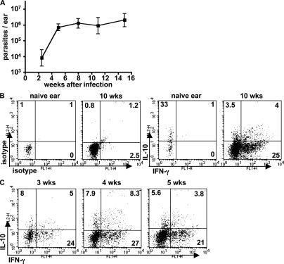
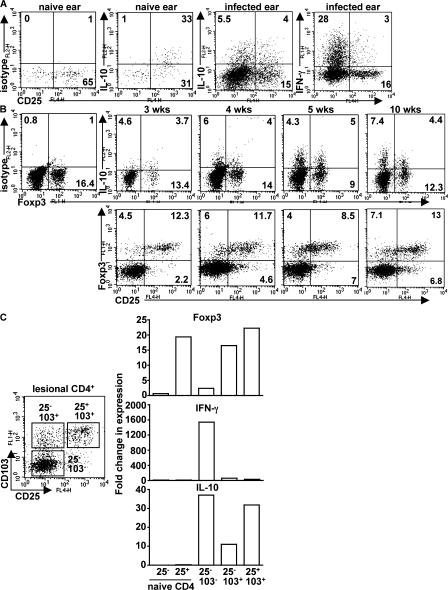
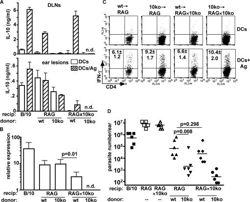
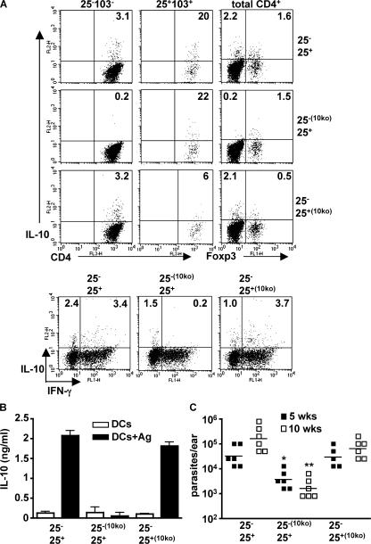
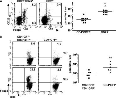
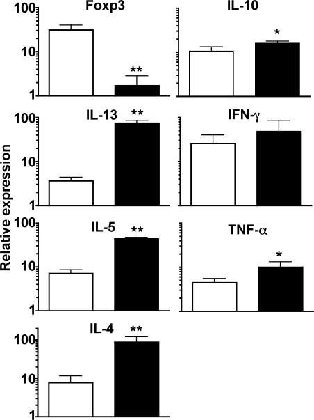
Nonhealing forms of leishmaniasis in humans are commonly associated with elevated levels of the deactivating cytokine IL-10, and in the mouse, normally chronic infections can be cleared in the absence of IL-10. Using a Leishmania major strain that produces nonhealing dermal lesions in a T helper type 1 (Th1) cell-polarized setting, we have analyzed the cellular sources of IL-10 and their relative contribution to immune suppression. IL-10 was produced by innate cells, as well as CD4(+)CD25(+)Foxp3(+) and CD4(+)CD25(-)Foxp3(-) T cells in the chronic lesion. Nonetheless, only IL-10 production by antigen-specific CD4(+)CD25(-)Foxp3(-) T cells, the majority of which also produced IFN-gamma, was necessary for suppression of acquired immunity in Rag(-/-) reconstituted mice. Surprisingly, Rag(-/-) mice reconstituted with naive CD4(+) T cells depleted of natural T regulatory cells developed more severe infections, associated with elevated levels of IL-10 and, especially, Th2 cytokines in the site. The data demonstrate that IL-10-producing Th1 cells, activated early in a strong inflammatory setting as a mechanism of feedback control, are the principal mediators of T cell-derived IL-10-dependent immune suppression in a chronic intracellular infection.


Figures

Figure 1.
Figure 1.
CD4+ T lymphocytes producing both IFN-γ and IL-10 are associated with nonhealing lesions in C57BL/6 mice infected with L. major NIH/Sd. Mice were inoculated intradermally with 1,000 metacyclic-stage parasites in each ear. (A) Parasite burdens in the ear lesions were monitored throughout the infection. Values represent the mean ± SD of six mouse ears at each time point during one experiment and are representative of at least three independent experiments. (B) 10 wk after infection, ear lesion cells or naive control ear cells were stimulated in vitro with PMA and ionomycin for 4 h and analyzed for intracellular cytokine production. (C) Ear lesion cells were analyzed for cytokine production at the indicated time points after infection. All cells shown are TCRβ+CD4+ lymphocytes, and quadrant values are the percentage of total gated CD4+ T cells. The plots are from six pooled ears per time point from one experiment and are representative of at least three.
Figure 2.
Figure 2.
CD4+ T cells producing IL-10 in response to infection with L. major NIH/Sd comprise multiple subsets. (A) Naive ear cells or cells from the chronic ear lesion were stimulated in vitro with PMA and ionomycin for 4 h and analyzed for intracellular cytokine production. (B) A kinetic analysis of IL-10 production and Foxp3 expression was performed on in vitro–restimulated lesional cells at the indicated time points. The data shown are TCRβ+CD4+ cells from six pooled ears from one experiment and are representative of at least three independent experiments. Quadrant values are the percentage of total gated CD4+ T cells. (C) 10 wk after infection, TCRβ+CD4+ ear lesion cells were purified by cell sorting into three subsets according to cell surface expression of CD25 and CD103. Real-time PCR analysis was performed to quantify gene expression of Foxp3, IFN-γ, and IL-10. Data are expressed as the fold change relative to naive CD4+CD25 lymph node cells. Two independent experiments were performed with similar results.
Figure 3.
Figure 3.
T cells are the dominant source of IL-10–mediated immune suppression in L. major NIH/Sd infection. 2 × 10⁶ T lymphocytes from lymph nodes of naive C57BL/10 or IL-10−/− mice were adoptively transferred into RAG−/− or RAG−/− × IL-10−/− recipients, followed by intradermal inoculation of L. major NIH/Sd 1 d later. 11 wk after infection, mice were killed for analysis. (A) Draining lymph node cells or ear lesion cells were stimulated in vitro with BMDCs in the absence or presence of L. major antigen for 72 h, followed by IL-10 measurement by ELISA. Groups are labeled with the source of donor T cells specified below either RAG−/− or RAG−/− × IL-10−/− recipient mice. Data are the mean ± SD of triplicate samples from one experiment. The experiment was performed three times with similar results. (B) Real-time PCR was performed on ear lesion tissue for quantification of IL-10. The values are the fold change in gene expression relative to naive controls. Data are the mean ± SD of four ears from one experiment and are representative of three experiments. (C) Ear lesion cells were stimulated in vitro with BMDCs or BMDCs infected with L. major amastigotes. After overnight incubation, monensin was added in the final 4 h, followed by measurement of intracellular IFN-γ. The plots shown are from one experiment, and the values in the upper left quadrants are the total number of IFN-γ+CD4+ cells/ ear × 10−3 and are the mean ± SD of three independent experiments. (D) Parasite burdens in the ear lesions were enumerated by limiting dilution. Unreconstituted RAG−/− and RAG−/− × IL-10−/− mice are also shown as controls. The data points are from six mice from one experiment and are representative of three independent experiments. Horizontal lines represent geometric means.
Figure 4.
Figure 4.
IL-10 produced by antigen-induced CD4+CD25Foxp3 T cells is necessary for the suppression of immunity. CD4+ lymph node T cells from naive WT or IL-10−/− mice were separated into CD25 and CD25+ fractions and adoptively cotransferred in a 10:1 ratio in the indicated combinations into RAG−/− recipients, followed by intradermal inoculation of 1,000 L. major NIH/Sd metacyclic promastigotes 1 d later. (A) 10 wk after infection, mice were killed, and lesion cells were stimulated with PMA and ionomycin for 4 h for measurement of intracellular IL-10 and IFN-γ. CD4+ cells were gated on CD25CD103 and CD25+CD103+ for identification of subsets of IL-10–producing cells. Foxp3 expression and IFN-γ/IL-10 was measured from total CD4+ cells. (B) Draining lymph node cells were stimulated in vitro with BMDCs in the absence or presence of L. major antigen for 72 h, followed by measurement of IL-10 by ELISA. Values shown are the mean ± SEM. (C) Parasite burdens in ear lesions were measured at 5 and 10 wk. The data points are from six mice in each group. The experiments were performed twice with similar results. Horizontal lines represent geometric means. *, P = 0.0005; **, P < 0.0001 in comparison to mice receiving CD25+/− T cells from IL-10–sufficient mice. Ag, antigen.
Figure 5.
Figure 5.
The absence of natural T reg cells exacerbates the infection. (A) 2 × 10⁶ naive CD4+CD25 T cells alone or in combination with 2 × 10⁵ CD4+CD25+ cells were adoptively transferred into RAG−/− recipients, followed by intradermal inoculation with L. major NIH/Sd 1 d later. 10 wk after infection, mice were killed for parasite burden and immune analysis. Surface CD25 and nuclear Foxp3 expression were measured in lesional cells, and the quadrant values represent the percentage of total CD4+ cells in the ear. For parasite burdens, data points are from 10 ears (from five mice) from one experiment and are representative of at least three independent experiments. Horizontal lines represent geometric means. *, P = 0.008. (B) 2 × 10⁶ CD4+GFP cells from naive Foxp3-GFP knock-in mice were adoptively transferred alone or in combination with 2 × 10⁵ CD4+GFP+ cells into RAG−/− recipients. 6 wk after infection, mice were killed, and lesions were analyzed for parasite burdens and cell composition. Foxp3 expression was measured in draining lymph nodes and ear lesions. Parasite burden data points are from six mice. The experiment was performed three times with similar results. Horizontal lines represent geometric means. **, P = 0.014.
Figure 6.
Figure 6.
Natural T reg cells suppress Th2 cell development. 2 × 10⁶ CD4+GFP cells from naive Foxp3-GFP knock-in mice were adoptively transferred alone (shaded bars) or in combination with 2 × 10⁵ CD4+GFP+ cells (open bars), followed by infection 1 d later. 6 wk after infection, mice were killed, and real-time PCR was performed on RNA isolated from ear lesions. Values shown are the mean ± SEM and are relative gene expression normalized to 18S rRNA. Data were generated from cells pooled from six mice in each group and are representative of three experiments. *, P < 0.05; **, P < 0.01.

References

    1. Sacks, D., and N. Noben-Trauth. 2002. The immunology of susceptibility and resistance to Leishmania major in mice. Nat. Rev. Immunol. 2:845–858. - PubMed
    1. Melby, P.C., F.J. Andrade-Narvaez, B.J. Darnell, G. Valencia-Pacheco, V.V. Tryon, and A. Palomo-Cetina. 1994. Increased expression of proinflammatory cytokines in chronic lesions of human cutaneous leishmaniasis. Infect. Immun. 62:837–842. - PMC - PubMed
    1. Louzir, H., P.C. Melby, A. Ben Salah, H. Marrakchi, K. Aoun, R. Ben Ismail, and K. Dellagi. 1998. Immunologic determinants of disease evolution in localized cutaneous leishmaniasis due to Leishmania major. J. Infect. Dis. 177:1687–1695. - PubMed
    1. Gasim, S., A.M. Elhassan, E.A. Khalil, A. Ismail, A.M. Kadaru, A. Kharazmi, and T.G. Theander. 1998. High levels of plasma IL-10 and expression of IL-10 by keratinocytes during visceral leishmaniasis predict subsequent development of post-kala-azar dermal leishmaniasis. Clin. Exp. Immunol. 111:64–69. (published erratum appears in Clin. Exp. Immunol. 1998. 112:547) - PMC - PubMed
    1. Ismail, A., A.M. El Hassan, K. Kemp, S. Gasim, A.E. Kadaru, T. Moller, A. Kharazmi, and T.G. Theander. 1999. Immunopathology of post kala-azar dermal leishmaniasis (PKDL): T-cell phenotypes and cytokine profile. J. Pathol. 189:615–622. - PubMed

Publication types