. 2007 Feb 13;104(7):2289-94.
doi: 10.1073/pnas.0611167104. Epub 2007 Feb 5.

Death-effector domain-containing protein DEDD is an inhibitor of mitotic Cdk1/cyclin B1

Affiliations

Death-effector domain-containing protein DEDD is an inhibitor of mitotic Cdk1/cyclin B1

Satoko Arai et al. Proc Natl Acad Sci U S A. .

Abstract

Accumulating evidence has shown that many molecules, including some cyclin-dependent kinases (Cdks) and cyclins, as well as the death-effector domain (DED)-containing FADD, function for both apoptosis and cell cycle. Here we identified that DEDD, which also possesses the DED domain, acts as a novel inhibitor of the mitotic Cdk1/cyclin B1 complex. DEDD associates with mitotic Cdk1/cyclin B1 complexes via direct binding to cyclin B1 and reduces their function. In agreement, kinase activity of nuclear Cdk1/cyclin B1 in DEDD-null (DEDD-/-) embryonic fibroblasts is increased compared with that in DEDD+/+ cells, which results in accelerated mitotic progression, thus exhibiting a shortened G2/M stage. Interestingly, DEDD-/- cells also demonstrated decreased G1 duration, which perhaps enhanced the overall reduction in rRNA amounts and cell volume, primarily caused by the rapid termination of rRNA synthesis before cell division. Likewise, DEDD-/- mice show decreased body and organ weights relative to DEDD+/+ mice. Thus, DEDD is an impeder of cell mitosis, and its absence critically influences cell and body size via modulation of rRNA synthesis.


Conflict of interest statement

The authors declare no conflict of interest.

Figures

Fig. 1.
Fig. 1.
Accelerated mitotic progression in DEDD−/− MEF cells. (A) Proliferation of 3T3 MEF cells. Cell numbers are plotted each day after the initial plating of 105 cells in a 10-cm-diameter plate. WT, DEDD+/+; KO, DEDD−/−; KO+DEDD 1 and 2, two clones of DEDD−/− cells transfected with DEDD. Data are averages of triplicates. Error bars indicate SEM. (B) BrdU incorporation analysis. (Upper) BrdU/7-amino-actinomycin D FACS profiles of WT, KO, and KO+DEDD cells. (Lower) First three lanes, various types of 3T3-MEF cells; last two lanes, 293T cells with or without DEDD overexpression.
Fig. 2.
Fig. 2.
DEDD associates with the Cdk1/cyclin B1 complex. (A) 293T cells expressing Flag-DEDD were synchronized and cytostained for DEDD (green) and 7-amino-actinomycin D (red) at indicated cell cycle phases. Subphase during mitosis was judged by the morphology of cells and DNA (chromosome). Non-TF, nontransfected 293 T cells; Int, interphase; Pro, prophase; Pro-meta, prometaphase. (B) In vivo binding assay. Flag-DEDD protein was coimmunoprecipitated (IP) with endogenous Cdk1/cyclin B1 from Flag-DEDD transfected 293T cells synchronized at G2/M (second lane) or at metaphase (third lane). As a control, nontransfected 293 cells at the G2/M phase (first lane) were analyzed. Cells at metaphase were also used for immunoprecipitation using an anti-cyclin B1 antibody (fourth lane) or a control rabbit IgG (fifth lane). Precipitates were analyzed by Western blotting using antibodies against Flag (FL), cyclin B1 (CyB1), or Cdk1. (C) Association of endogenous DEDD with Cdk1/cyclin B1 complexes. Precipitates by an anti-Cdk1 antibody from cell lysates of wild-type (nontransfected) 293T cells synchronized at G2/M (second lane), postmitotic G1 (third lane), or metaphase (fourth lane) were tested by Western blotting using antibodies against DEDD (Top), cyclin B1 (Middle), or Cdk1 (Bottom). As a control, asynchronous 293T cells were analyzed (first lane). Precipitates with a control rabbit IgG from cells at metaphase were also Western-blotted (fifth lane). The anti-human and anti-mouse DEDD polyclonal antiserum was newly generated by immunizing rabbits with a KLH-conjugated DEDD peptide (CPDLVDKYLEETSIRYVT). (D) Pan-caspase inhibitor z-VAD does not inhibit Cdk1/cyclin B1/DEDD complex formation. 293T cells with (second and third lanes) or without (first lane) Flag-DEDD transfection were arrested at metaphase. A proportion of transfected cells were also incubated with 50 μM of z-VAD-fmk (Biovision) for the last hour (third lane). Cdk1/cyclin B1/DEDD complexes were immunoprecipitated by using an anti-Flag antibody. Precipitates were analyzed by Western blotting for Cdk1 (Upper) or Flag-DEDD (Lower). (E) In vitro binding assay using purified Flag-tagged DEDD, HA-tagged Cdk1, and GST-cyclin B1 fusion protein. A prey protein was pulled down by either type of bait protein. As controls, prey proteins were also incubated in the absence of the respective bait protein, followed by precipitation (Bait: −). Precipitates were analyzed by Western blotting (WB) using antibodies against Flag (FL; for DEDD), HA (for Cdk1), or GST (for cyclin B1). (F Top) Schematic showing of the structure of F-DEDD, N-DEDD, and C-DEDD. Light blue region, nuclear localizing sequences; black region, DED domain; orange region, proline-rich region. Numbers indicate amino acids starting from the first methionine. (F Middle and Bottom) Binding assay of the F, N, and C-DEDD to GST-cyclin B1. Flag-tagged DEDD variant proteins (baits) were purified from 293 T cells overexpressing the respective proteins and incubated with GST-cyclin B1 (prey), followed by a pull-down. Precipitates were analyzed by Western blotting, either for binding using an anti-GST antibody (Middle) or for bait proteins using an anti-Flag (FL) antibody (Bottom). As a control, the prey protein, GST-cyclin B1, was also loaded on the gel and Western-blotted with the anti-GST antibody (WB: GST, fifth lane). The N-DEDD protein appeared to yield dimers due to homotypic interactions (31) (indicated by an arrowhead in Bottom). (G) FADD does not associate with the Cdk1/cyclin B1/DEDD complex. The Cdk1/cyclin B1/DEDD complex was immunoprecipitated from nocodazole-treated 293T cells with (second lane) or without (first lane) Flag-DEDD transfection by using an anti-Flag antibody. Precipitates were analyzed by Western blotting for FADD (first panel), Cdk1 (second panel), or Flag-DEDD (third panel).
Fig. 3.
Fig. 3.
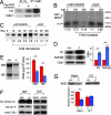
Association of DEDD reduces kinase activity of the Cdk1/cyclin B1 complex. (A) Kinase activity on the histone H1 substrate. Activities were normalized by the amount of precipitated Cdk1 (Western blotting; Lower). The negative control (first lane) revealed an invisible level of Cdk1 but a background level of activity. (B) DEDD-free Cdk1/cyclin B1 was immunopurified from DEDD−/− MEF cells by using an anti-cyclin B1 antibody. The kinase activity of the precipitate on the histone H1 substrate was assayed in the presence of different amounts of GST-DEDD (0, 100, and 200 ng) or GST (100 and 200 ng) protein. Fold increase indexes compared with the signal that was obtained without GST-DEDD or GST protein are displayed. The intensities of the signals were quantified by using image analysis software (NIH Image 1.63). No phosphorylation of GST-DEDD (60 kDa, size indicated) was detected. (C) Addition of recombinant GST-DEDD stimulates Pol.I transcription in vitro. Comparable amounts of GST-DEDD (second to fifth lanes) or GST alone (sixth to eighth lanes) were added to a fractionated murine nuclear extract (DEAE-280) from DEDD−/− MEF cells containing Cdk1/cyclin B1 complexes, and Pol.I transcription was initiated in the presence of nucleotides and linearized template DNA (34, 35). Radiolabeled runoff transcripts were separated on polyacrylamide gels and quantified by using a PhosphorImager. Fold stimulation indexes compared with the signal that was obtained without GST-DEDD or GST protein are displayed. Three independent experiments were performed and revealed similar results. (D Left) Kinase activity assay of Cdk1/cyclin B1 precipitated from DEDD+/+ (+/+) or DEDD−/− (−/−) 3T3 MEF cell nuclei using histone H1 (Top) or TAFI95 as substrate (Middle). Western blotting for the precipitated Cdk1 is shown in Bottom. (D Right) The relative activities in −/− cells (blue bars) to those in +/+ cells (red bars) are presented. Data are averages ± SEM of three independent assays. (E) The rRNA levels. (Left) Total RNA from 5 × 10⁵ DEDD+/+ (+/+) and DEDD−/− (−/−) 3T3 MEF cells was separated on an agarose gel. (E Right) Absolute amounts of 28S and 18S rRNA per 106 cells. Means ± SEM are presented. (F) Phosphorylation status of Cdk1. Cdk1 was immunoprecipitated either from DEDD+/+ (+/+) or DEDD−/− (−/−) 3T3 MEF cells (first two lanes), 293T or 293T cells with DEDD transfection (293+DEDD) (third and fourth lanes), and the phosphorylation of the Thr-14/Tyr-15 (Top) and the Thr-161 (Middle) residues were analyzed by Western blotting. Data were normalized to the precipitated Cdk1 amounts (Bottom). (G) The pre-rRNA synthesis. DEDD+/+ (+/+) and DEDD−/− (−/−) 3T3 MEF cells were enriched at G1 by serum starvation. Total RNA isolated from 5 × 10⁵ G1-enriched cells, as well as from asynchronous cells as a control, was analyzed for 45S pre-rRNA by Northern blotting by using a mouse rDNA (from −168 to + 158) fragment as a probe (Upper). The amount of 45S pre-rRNA was also analyzed by quantitative RT-PCR. Data are described as relative amounts of 45S pre-rRNA normalized to 36B4 mRNA (Lower). Means ± SEM of three independent analyses are presented.
Fig. 4.
Fig. 4.
Lack of DEDD reduces size in cells and mice. (A Left and Center) Representative FSC/SSC profiles of DEDD+/+ (WT) and DEDD−/− (KO) MEF cells. (A Right) The FSC and SSC of DEDD−/− cells (blue bars) relative to those of DEDD+/+ (red bars) cells. (B) The FSC and SSC of DEDD−/− cells transfected with DEDD (green bars) relative to those of DEDD−/− (blue bars) cells. Data are averages from three different lines for each type of cell. Error bars indicate SEM. (C) Mouse photo showing DEDD+/+ (on the top) and DEDD−/− (on the bottom) mice. Both mice were 6 weeks of age and had been backcrossed to B6 for 10 generations. (D) Body weights. Five mice per group were analyzed, and the averages are presented. All mice used were littermates obtained from intercross-breeding of DEDD+/− mice that had been backcrossed to B6 mice for nine generations. Error bars indicate SEM. The differences between WT and KO mice were statistically significant in both males (M) and females (F) (Mann–Whitney test; P < 0.01). (E) Weights of indicated organs. Lung(rt), right lobes of the lung; red bars, DEDD+/+; blue bars, DEDD−/−. Data are averages from five male mice.

Similar articles

Cited by

References

    1. Jorgensen P, Nishikawa JL, Breitkreutz BJ, Tyers M. Science. 2002;297:395–400. - PubMed
    1. Jorgensen P, Rupes I, Sharom JR, Schneper L, Broach JR, Tyers M. Genes Dev. 2004;18:2491–2505. - PMC - PubMed
    1. Jorgensen P, Tyers M. Curr Biol. 2004;14:R1014–R1027. - PubMed
    1. Kellogg DR. J Cell Sci. 2003;116:4883–4890. - PubMed
    1. Rupes I. Trends Genet. 2002;18:479–485. - PubMed

Publication types

MeSH terms