. 2007 Feb 13;104(7):2283-8.
doi: 10.1073/pnas.0610877104. Epub 2007 Feb 5.

Targeted cell killing by reconstituted caspases

Affiliations

Targeted cell killing by reconstituted caspases

Dattananda S Chelur et al. Proc Natl Acad Sci U S A. .

Erratum in

  • Proc Natl Acad Sci U S A. 2008 Jan 15;105(2):824

Abstract

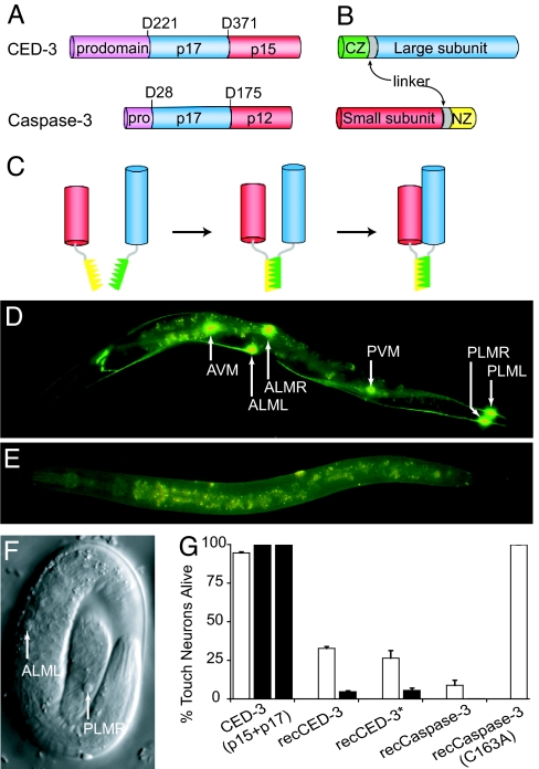
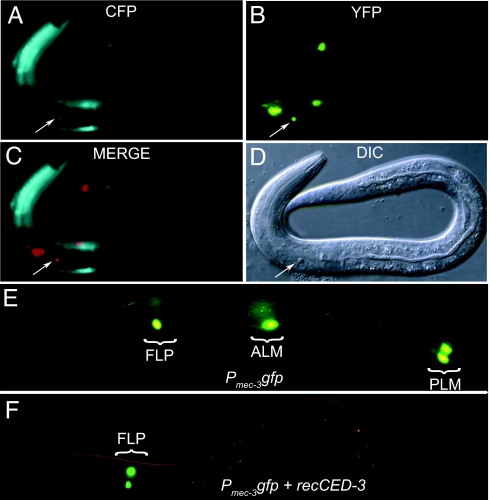
We have developed a two-component system involving reconstituted caspase (recCaspase) for selective and/or conditional ablation of targeted cells. Caspases, the executioners of programmed cell death, are normally synthesized as inactive zymogens and are activated by proteolytic processing of their subunits. We show here, using two different caspases, Caenorhabditis elegans CED-3 and human Caspase-3, that coexpression of the subunits generates constitutively active caspase activity that leads to cell death. This recCaspase activity, however, occurred only when the subunits associated through binding of linked antiparallel leucine-zipper domains. We exploited the dual-component nature of recCaspases by expressing the individual subunits from combinations of promoters either to target selectively the subset of cells for apoptosis or induce cell death in specific cells at specific times during development. The high degree of target specificity and tight regulation of induction of recCaspase would be advantageous in creating animal models that are ablated for specific cells and in other targeted cell killings.


Conflict of interest statement

Conflict of interest statement: D.S.C. has submitted a patent based on this work.

Figures

Fig. 1.
Fig. 1.
Cell killing activity of recCaspases in C. elegans. (AC) Schematics of reconstitution of activated caspase from its subunits. (A) Subunit organization in the CED-3 and caspase-3 procaspases. The aspartate residues that are cleaved for activation are shown. (B) recCaspase components. Antiparallel leucine-zipper domains NZ (yellow) and CZ (green) are attached to the small (red) and large (blue) caspase subunits through a linker sequence (gray). (C) Formation of recCaspase. Interaction between the leucine-zipper domains leads to oligomerization of the caspase subunits that, in turn, results in the association and activation of caspase subunits. The precise subunit composition of the final recCaspase is not known. (DG) Cell killing by recCaspases in C. elegans. Fluorescent images of adult worms expressing either the CED-3 subunits without the leucine-zipper sequences (p17 + p15) (D) or the recCED-3 subunits from touch neuron-specific mec-18 promoter (E). The GFP-positive touch receptor neurons are indicated by arrows. (F) Differential interference contrast (DIC) image of an embryo showing recCED-3 mediated apoptosis of an ALM and a PLM neuron. (G) Percentage of touch receptor neurons remaining in adults expressing the CED-3 subunits (p15 + p17) and recCaspases. GFP-positive cells were counted in 1-day-old adults expressing recCaspase from stable line with extragenic arrays of the constructs (open bars) or from the chromosomally integrated arrays (filled bars). Three stable lines or three different experiments in case of integrated lines were used to calculate mean and SEM. recCED-3* contains an activating mutation (45).
Fig. 2.
Fig. 2.
recCaspase-3 activity in HeLa cells. (AC) Induction of recCaspase-3 activity with 1 μg/ml doxycycline. (AC Left) Fluorescent micrographs. (AC Right) DIC photomicrograph of the same field. Without doxycycline (A), the caspase3-sensor EYFP is found throughout cells; after induction (B and C), EYFP fluorescence localizes to the nucleus. (C) Dying cells (arrows) exhibiting the characteristic apoptotic morphology. (D) recCaspase-3, but not caspase-3, subunits are active in HeLa cells. Percentages of nuclear-localized GFP cells out of the total GFP-positive cells are given. Observations from three independent experiments were used to calculate mean and SEM.
Fig. 3.
Fig. 3.
Targeted cell killing by recCaspase expressed from a combination of promoters with overlapping expression patterns. (AD) Targeting the AVD interneuron (indicated by arrow) for cell death by recCaspase3 expressed from a combination of promoters Pnmr-1cz::caspase-3(p17) and Pcfi-1caspase-3(p12)::nz. Although the cfi-1 promoter expresses CFP in many neurons and head muscle (A) and the nmr-1 promoter expresses yellow fluorescent protein in multiple neurons (B), they are both expressed only in the AVD cells (C; the yellow fluorescent protein image was artificially colored red). (D) The death of an AVD neuron (arrow) as seen by DIC microscopy. (E and F) Restriction of GFP to FLP neurons by recCED-3 mediated cell death. (E) Without recCED-3, Pmec-3gfp is expressed in the ALM and PLM touch neurons and the FLP neurons of a newly hatched larva. (F) When recCED-3 is expressed by using Pmec-3 cz::ced-3(p17) and Pmec-18ced-3(p15)::nz, the touch neurons, which express both the promoters, die, and GFP fluorescence is found only in the FLP neurons.
Fig. 4.
Fig. 4.
Conditional induction of recCaspase-3 activity in muscle cells. (A) Expression of only the p17 subunit of the recCaspase-3 from the heat shock promoter [(Phsp-16 cz::caspase-3(p17)] had no effect on survival of body wall muscle. (A and B) GFP fluorescence from the muscle-specific myo-3 promoter (A and B Upper); a DIC image of the same animal (A and B Lower). For both animals, newly hatched L1 larvae were heat shocked for two hours and photographed 24 h later. (B) Expression of Pmyo-3 caspase-3(p12)::nz (causing the p12 subunit of recCaspase to be expressed in muscles) in combination with Phsp-16 cz::caspase-3(p17) resulted in death of the body wall muscles. (C) Enlargement of the DIC image of the animal in B showing the vacuolated deaths of muscle cells. (D) Superimposed DIC and fluorescence (Pmyo-3gfp) images of an embryo heat shocked for 30 min and photographed 8 h later. Arrows indicate some of the apoptotic nuclei, which show the characteristic refractile, flat disk-like morphology. (E) Occurrence of muscle cell death with induction of recCaspase activity at different times. Animals were heat shocked at the indicated times and scored 48 h later for paralysis caused by the death of muscle cells. Data from the expression of the single subunit (from Phsp-16; green line) as well as both the subunits (from Phsp-16 and Pmyo-3; blue line) of recCaspase are shown. Data from at least two stable lines (50 animals per line) and three independent experiments were used to calculate mean and SEM. (F) Time course of induction of recCaspase activity. First-stage larvae expressing both the subunits of the recCaspase were collected 12 h after hatching and subjected to a 2-h heat shock. At the indicated times after heat shock, animals were scored as completely paralyzed (blue), partially paralyzed (red), or wild type. Data from three stable lines and at least 100 animals per line were used to calculate mean and SEM. The mean values of partially and completely paralyzed animals were added to obtain the total number of animals affected (black) at each time point.

Similar articles

Cited by

References

    1. Sulston JE, White JG. Dev Biol. 1980;78:577–597. - PubMed
    1. Bernhardt RR, Nguyen N, Kuwada JY. Neuron. 1992;8:869–882. - PubMed
    1. Shah EM, Jay DG. Curr Opin Neurobiol. 1993;3:738–742. - PubMed
    1. Palmiter RD, Behringer RR, Quaife CJ, Maxwell F, Maxwell IH, Brinster RL. Cell. 1987;50:435–443. - PubMed
    1. Borrelli E, Heyman R, Hsi M, Evans RM. Proc Natl Acad Sci USA. 1988;85:7572–7576. - PMC - PubMed

Publication types