. 2007 Feb 13;104(7):2247-52.
doi: 10.1073/pnas.0607238104. Epub 2007 Feb 5.

X-ray fluorescence microscopy reveals large-scale relocalization and extracellular translocation of cellular copper during angiogenesis

Affiliations

X-ray fluorescence microscopy reveals large-scale relocalization and extracellular translocation of cellular copper during angiogenesis

Lydia Finney et al. Proc Natl Acad Sci U S A. .

Abstract

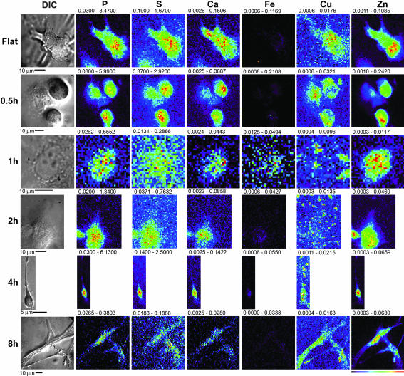
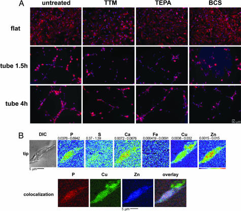
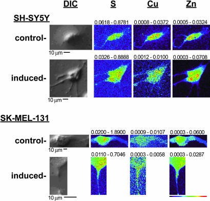
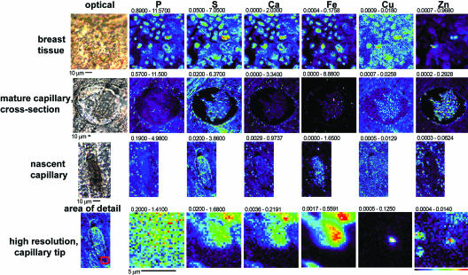
Although copper has been reported to influence numerous proteins known to be important for angiogenesis, the enhanced sensitivity of this developmental process to copper bioavailability has remained an enigma, because copper metalloproteins are prevalent and essential throughout all cells. Recent developments in x-ray optics at third-generation synchrotron sources have provided a resource for highly sensitive visualization and quantitation of metalloproteins in biological samples. Here, we report the application of x-ray fluorescence microscopy (XFM) toin vitro models of angiogenesis and neurogenesis, revealing a surprisingly dramatic spatial relocalization specific to capillary formation of 80-90% of endogenous cellular copper stores from intracellular compartments to the tips of nascent endothelial cell filopodia and across the cell membrane. Although copper chelation had no effect on process formation, an almost complete ablation of network formation was observed. XFM of highly vascularized ductal carcinomas showed copper clustering in putative neoangiogenic areas. This use of XFM for the study of a dynamic developmental process not only sheds light on the copper requirement for endothelial tube formation but highlights the value of synchrotron-based facilities in biological research.


Conflict of interest statement

The authors declare no conflict of interest.

Figures

Fig. 1.
Fig. 1.
XFM scans of HMVECs undergoing tubulogenesis. Silicon nitride windows were coated with either a thin layer of Matrigel or a layer of gelatin. HMVECs were plated on these substrates and exposed to VEGF and bFGF. The process was stopped at various times subsequent to initiation; the cells were then fixed, and both light microscope and XFM images were obtained. The scans are representative of the five cells scanned at each time point. The scale of the scans is shown below each image, and a color table is shown to the bottom right. The maximum and minimum threshold values in micrograms per squared centimeter are given above each frame. Scans were obtained by using 10.0-keV incident energy with dwell times of 1 sec per pixel and 1-μm steps through the sample.
Fig. 2.
Fig. 2.
Role of copper in HMVEC filopodia extension. (A) HMVECs were plated in either gelatin- or Matrigel-coated dishes, stimulated with bFGF and VEGF, and untreated or exposed to 100 μM TEPA, 100 μM TTM, or 100 μM BCS. The cells were incubated for either 1.5 or 4 h, fixed and permeabilized, and stained with Hoescht 33342 and Alexa Fluor 660–phalloidin. Cells were then imaged, and the Hoescht signal and phalloidin signal were pseudocolored blue and red, respectively. (B) Areas at the tips of HMVEC filopodia extensions were scanned by XFM at high resolution. (Upper) The optical image and metal maps. (Lower) False-color images of P, Zn, and Cu and the overlay of these images.
Fig. 3.
Fig. 3.
XFM scans of cells undergoing neural process extension. SH-SY5Y cells were applied to either gelatin- or Matrigel-coated silicon nitride windows and exposed to bFGF and IGF for 3 d. SK-MEL-131 cells were plated directly on silicon nitride windows and either untreated or exposed to 45 μM genistein for 2 d. The cells were fixed and washed, optical images were acquired, and the cells were scanned by XFM analysis. Representative output is shown, with the scale bar and color table shown below the scans. The maximum and minimum threshold values in micrograms per squared centimeter are given above each frame.
Fig. 4.
Fig. 4.
XFM scans of human breast-infiltrating ductal carcinoma tumor tissues. Areas of breast and endothelial cells were identified in paraffin-embedded tissue, and XFM scans (1–2 sec per pixel) of such areas were performed. For each representative scan, an optical image is shown to the left. Maps of each metal show areas of lowest content to highest content scaled to a rainbow color scale shown to the bottom right. The size scale of each scan is shown by the scale bar under each scan, and the minimal and maximal content displayed in micrograms per squared centimeter is listed above each image. The region of the nascent capillary on the left from which the high-resolution scans to the right are derived is shown as a red square.

Similar articles

Cited by

References

    1. Lippard SJ, Berg JM. Principles of Bioinorganic Chemistry. Mill Valley, CA: Univ Sci Books; 1994.
    1. Valko M, Morris H, Cronin MT. Curr Med Chem. 2005;12:1161–1208. - PubMed
    1. O'Halloran TV, Culotta VC. J Biol Chem. 2000;275:25057–25060. - PubMed
    1. Finney LA, O'Halloran TV. Science. 2003;300:931–936. - PubMed
    1. Ziche M, Jones J, Gullino PM. J Natl Cancer Inst. 1982;69:475–482. - PubMed

Publication types