

doi: 10.1021/ja0693587.
Epub 2007 Feb 7.
Reactivation of the p53 tumor suppressor pathway by a stapled p53 peptide
Affiliations
- PMID: 17284038
- PMCID: PMC6333086
- DOI: 10.1021/ja0693587
Item in Clipboard
Reactivation of the p53 tumor suppressor pathway by a stapled p53 peptide
J Am Chem Soc.
.
Erratum in
- J Am Chem Soc. 2007 Apr 25;129(16):5298
No abstract available
Figures

Synthesis, sequence, and biochemical analysis of SAH-p53 peptides. (A) Non-natural amino acids were synthesized as described and cross-linked by ruthenium-catalyzed ring-closing olefin metathesis. (B) SAH-p53 compounds were generated by stapling the p5314-29 sequence at the indicated positions. Charge, α-helicity, hDM2 binding affinity, cell permeability, and impact on cell viability are indicated for each compound. (C, E) Circular dichroism spectra revealed enhancement of α-helicity for SAH-p53 compounds. (D, F) Fluorescence polarization assays using FITC-peptides and hDM217-125 demonstrated subnanomolar hDM2-binding affinities for select SAH-p53 peptides. Note: UAH-p53-8 is the “unstapled” form of SAH-p53-8.
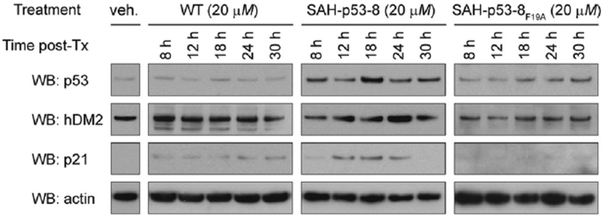
SAH-p53-8 reactivates the p53 transcriptional pathway. The hDM2 overexpressing SJSA-1 cells were exposed to the indicated peptides, and Western analyses for p53, hDM2, and p21 were performed at 8–30 h of treatment.
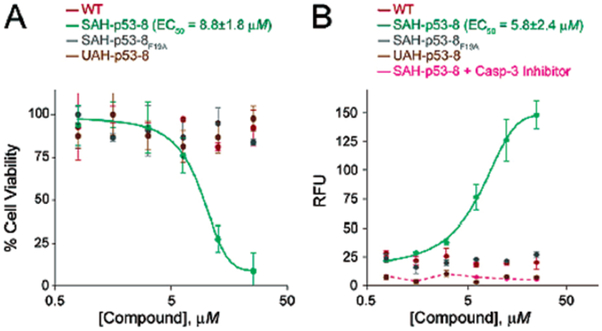
Reactivation of apoptosis in SAH-p53-8-treated SJSA-1 cells. SAH-p53-8 demonstrated specific, dose-dependent cytotoxicity and apoptosis induction as measured by (A) CellTiter-Glo (Promega) and (B) Caspase-3 activation assays (Oncogene).
References
Publication types
MeSH terms
Substances
Grants and funding
LinkOut - more resources
Full Text Sources
Other Literature Sources
Research Materials
Miscellaneous
