. 2007 Feb 6:2:1.
doi: 10.1186/1750-2187-2-1.

ArhGAP9, a novel MAP kinase docking protein, inhibits Erk and p38 activation through WW domain binding

Affiliations

ArhGAP9, a novel MAP kinase docking protein, inhibits Erk and p38 activation through WW domain binding

Boon K Ang et al. J Mol Signal. .

Abstract

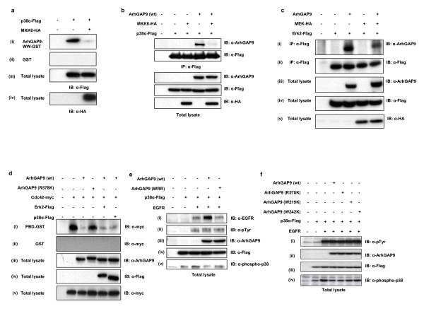
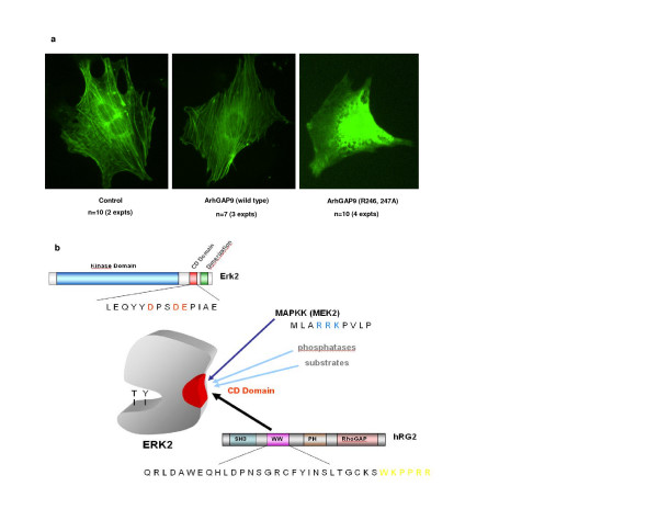
We have identified human ArhGAP9 as a novel MAP kinase docking protein that interacts with Erk2 and p38alpha through complementarily charged residues in the WW domain of ArhGAP9 and the CD domains of Erk2 and p38alpha. This interaction sequesters the MAP kinases in their inactive states through displacement of MAP kinase kinases targeting the same sites. While over-expression of wild type ArhGAP9 caused MAP kinase activation by the epidermal growth factor receptor (EGFR) to be suppressed and preserved the actin stress fibres in quiescent Swiss 3T3 fibroblasts, over-expression of an ArhGAP9 mutant defective in MAP kinase binding restored EGFR-induced MAP kinase activation and resulted in significant disruption of the stress fibres, consistent with the role of Erk activation in disassembly of actin stress fibres. The interaction between ArhGAP9 and the MAP kinases represents a novel mechanism of cross-talk between Rho GTPase and MAP kinase signaling.


Figures

Figure 1
Figure 1
Erk2 binds to human ArhGAP9 at the WW domain. a. Evolutionary conservation of ArhGAP9 and ArhGAP12. Schematic representation of ArhGAP9 and ArhGAP12 domain structures of different species as predicted by SMART domain prediction tool. b. Identification of Erk2 as an interacting protein of the WW domain of human ArhGAP9. Immobilized ArhGAP9-WW-GST precipitated several proteins (bands a-g) from rat brain lysate detected by Colloidal Coomassie Blue. The bands were excised for in-gel reduction, S-alkylation and trypsin hydrolysis. The peptides were identified by mass spectrometry. Bands f and g generated 15 unique peptides covering 40% of the MAP kinase, Erk2 sequence.
Figure 2
Figure 2
Erk2 and p38α, but not Jnk1 interacted with ArhGAP9 WW domain. a. Specificity of Erk2 and p38α binding to the WW domain of ArhGAP9. MAP kinases, Erk2-Flag, p38α-Flag or Jnk1-Flag, were transiently expressed in 293T cells and the lysates used for in vitro binding assay with (i) ArhGAP9-WW-GST or (ii) GST control, followed by western blotting with α-Flag. (iii) Total cell lysates were separated by SDS-PAGE and immunoblotted with α-Flag. b. Full length Erk2 and p38α interact with ArhGAP9 in vitro. Flag-tagged ArhGAP9 was expressed in 293T cells and the lysates were incubated with immobilized (i) Erk2-GST, (ii) p38α-GST or (iii) GST alone as a control. Bound Erk2 and p38α was detected by western blotting with α-Flag. (iv) Total lysates were immunoblotted with α-Flag. c. Interaction of ArhGAP9 with MAP kinases in vivo. ArhGAP9 and MAP kinases, Erk2-Flag or p38α-Flag were expressed in 293T cells. The lysates were immunoprecipitated with α-Flag, followed by western blotting with (i) α-ArhGAP9 or (ii) α-Flag. (iii, iv) Immunoblotting of total cell lysates with α-ArhGAP9 or α-Flag. d. Erk2 and p38α interact specifically with the WW domain of ArhGAP9 but not ArhGAP12 or Nedd4. MAP kinases Erk2-Flag, p38α-Flag or Jnk1-Flag was expressed in 293T cells and the lysates were incubated with GST fusion proteins of the WW domains of ArhGAP9, ArhGAP12 or Nedd4, or GST alone immobilized on glutathione Sepharose beads, as indicated in the figure. (i) Specifically bound proteins were detected by western blotting with α-Flag. (ii) Total cell lysates were immunoblotted with α-Flag. e. Erk2 and p38α do not interact with mouse ArhGAP9 in vitro. Flag-tagged Erk2, p38α or Jnk1 plasmid was expressed in 293T cells. Immobilized GST proteins for (i) WW domain of human ArhGAP9, (ii) N-terminal fragment of mouse ArhGAP9 (residues 1–350) or (iii) GST alone was incubated with the lysates. Bound Erk2, p38α or Jnk1 was detected by western blotting with α-Flag. (iv) Total cell lysates were immunoblotted with α-Flag. f. Erk2 interact with human but not mouse. Human or mouse ArhGAP9-Flag was expressed in 293T cells, individually or together with Erk2-HA. The lysates were immunoprecipitated with α-Flag. (i) Bound Erk2 was detected by western blotting with α-HA. (ii) Immunoprecipitated human or mouse ArhGAP9 was detected by α-Flag. Total cell lysates were immunoblotted with (iii) α-Flag or (iv) α-HA.
Figure 3
Figure 3
The ArhGAP9 is a novel MAP kinase docking protein. a. Alignment of the WW domains of ArhGAP9 and ArhGAP12. WW domains of human ArhGAP9 and ArhGAP12 were aligned using ClustalW, highlighted in yellow. The di-Arginine motif at the extreme C terminus of ArhGAP9 WW domain is highlighted in cyan. b. R246 and R247 in the WW domain of human ArhGAP9 are required for binding to MAP kinases. Flag-tagged Erk2, p38α or Jnk1 was expressed in 293T cells and the lysates were incubated with immobilized ArhGAP9-WW-GST, ArhGAP9-WW-(R246, 247A)-GST mutant, or GST alone as a control. Specifically MAP kinases were detected by western blotting with α-Flag. (ii) The expression levels of Erk2, p38α and Jnk1 were confirmed to be equivalent by immunoblotting of the total cell lysates with α-Flag. c. R246 and R247 are required for ArhGAP9 WW domain binding to CD domain of MAP kinase in vitro. Flag-tagged ArhGAP9 (wild type or mutants R246A, R247A or R246, 247A) was expressed in 293T cells and the lysates were incubated with the (i) Erk2-CD-GST or (ii) GST alone. Bound ArhGAP9 was detected by immunoblotting with α-Flag. (iii) Total cell lysates were immunoblotted with α-Flag. d. R246 and R247 are required for ArhGAP9 WW domain binding to MAP kinase in vivo. Flag-tagged Erk2 or p38α was expressed individually or together with full-length ArhGAP9 [wildtype or the R246, 247A mutant (RR)] in 293T cells, as indicated in the figure. The lysates were immunoprecipitated with α-Flag followed by western blotting of the immunocomplexes with (i) α-ArhGAP9 or (ii) α-Flag. Total cell lysates were immunoblotted with (iii) α-ArhGAP9 or (iv) α-Flag. e. K243, P244 and P245 within the WW domain of ArhGAP9 are also important for MAP kinase binding. Flag-tagged Erk2 were transiently expressed either individually or together with full-length ArhGAP9 [wildtype or K243A, P244A and P245A mutants] in 293T cells. The lysates were then subjected to immunoprecipitation with α-Flag. The immunocomplexes were resolved by SDS-PAGE followed by western blotting with (i) α-ArhGAP9 or (ii) α-Flag. Total cell lysates were separated by SDS-PAGE and immunoblotted with (iii) α-ArhGAP9 or (iv) α-Flag.
Figure 4
Figure 4
The Common Docking (CD) domain of MAP kinase mediated the binding to the WW domain of ArhGAP9. a. Flag-tagged full-length ArhGAP9 (wild type or the R246,247A mutant, RR) was expressed 293T cells and the lysates were incubated with GST fusion of (i) the CD domain of Erk2 (Erk2-CD-GST, residues 300–358), (ii) a fragment of Erk2 deleted of the CD domain (Erk2-ΔCD-GST, residues 1–300) or (iii) GST alone as a control. Bound ArhGAP9 was detected by western blotting with α-Flag. (iv) Total cell lysates were immunoblotted with α-Flag. b. Acidic residues in the CD domain of MAP kinases are important for interaction with ArhGAP9. (i) Alignment of the Common Docking (CD) domains of Erk2, p38α and Jnk1, the acidic residues mutated to alanine are indicated in superscript. Flag-tagged Erk2 (wild type or D316A, D319A or E320A mutants) or p38α (wild type or D312A, D315A or E316A mutants) was expressed 293T cells. The lysates were incubated with (ii) ArhGAP9-WW-GST or (iii) GST alone. Bound Erk2 or p38α was detected by western blotting with α-Flag. (iv) Total cell lysates were immunoblotted with α-Flag. c. The activation loop of MAP kinase was not involved in binding with ArhGAP9. Flag-tagged Erk2 (wild type or T183, Y185A mutant) and p38α (wild type or T180, Y182A mutant) were expressed in 293T cells. The lysates were incubated with immobilized (i) ArhGAP9-WW-GST or (ii) GST alone. Bound Erk2 or p38α was detected by western blotting with α-Flag. (iii) Total cell lysates were immunoblotted with α-Flag. d. Far-UV CD spectra of WW of ArhGAP9 in complex with MAP kinase CD domain peptides. (i) The CD spectra of the WW domain of ArhGAP9 indicating that the protein folded properly and has mostly β-sheets and random coils. (ii-iv) CD spectra of ArhGAP9 WW domain in complex with Jnk1, p38α and Erk2 peptides, respectively. Conformational changes were observed for the case of ArhGAP9 WW domain in complex with Erk2 and p38α peptides. As for Jnk1, little effect in the spectra profile was observed when compared with WW domain alone, indicating that no significant binding of the Jnk1 peptide had occurred. e. Structural alignment of the ArhGAP9-binding regions in the CD domains of Erk2, p38α and Jnk1. (i) A stereo view of structural alignment of Erk2 (red) with p38α (maroon) (ii) A stereo view of structural alignment of Erk2 with Jnk1. The residues are shown in stick model and the key mutated residues of Erk2 are marked.
Figure 5
Figure 5
ArhGAP competes with MAP kinase kinases (MAPKKs) to suppress MAP kinase activation. a. Coexpression of MKK6 dimished the binding of p38α to the WW domain of ArhGAP9. p38α-Flag was transfected alone or together with HA-tagged activated mutant of MKK6 in 293T cells and the lysates were incubated with (i) ArhGAP9-WW-GST or (ii) GST alone as a control. Bound p38α was detected by western blotting with α-Flag. (iii, iv) Total cell lysates were immunoblotted with α-Flag or α-HA, respectively. b. Overexpression of MKK6 diminished the binding between ArhGAP9 and p38α. Flag-tagged p38α, HA-tagged MKK6 and ArhGAP9 were expressed in 293T cells as indicated in the figure. The immunocomplex of p38α was analyzed by western blotting with (i) α-ArhGAP9 and (ii) α-Flag. (iii-v) Total lysates were immunoblotted with α-ArhAGAP9, α-Flag and α-HA, respectively. c. Reduction of ArhGAP9 binding to Erk2 by MEKoverexpression in vivo. Flag-tagged Erk2, HA-tagged MEK2 and ArhGAP9 were transiently transfected in 293T cells as indicated in the figure. Immunoprecipitation was carried out with α-ArhGAP9, followed by western blotting with (i) α-ArhGAP9 α-Flag and (ii) α-HA. Western blotting of the total lysates with (iii) α-phospho-Erk showed that Erk2 was activated when coexpressed with MEK2. Western blotting of the total lysates with (iv) α-ArhAGP9, (v) α-Flag and (vi) α-HA showed the expression of ArhGAP9, Erk2 and MEK2 in the total cell lysates. d. MAP kinase binding has no significant effect on ArhGAP9 RhoGAP activity. ArhGAP9 (wild type or the GAP-inactive mutant, R578K) was transfected with myc-tagged Cdc42, as well as Flag-tagged Erk2 or p38α in 293T cells, as indicated in the figure. (i) The lysates were incubated with immobilized PBD-GST followed by detection of the relative amounts of active cdc42 precipitated by western blotting with α-myc. (ii) GST alone was used as a control. (iii-v) Total cell lysates were immunoblotted with α-ArhGAP, α-Flag or α-myc, rspectively. e. ArhGAP9 binding suppresses MAP kinase activation by EGFR. ArhGAP9 [wild type or the triple mutant W242K, R246A, R247A (WRR)] was expressed in 293T cells alone or with EGFR and Flag-tagged p38α, as indicated in the figure. (i-v) Total cell lysates were immunoblotted with antibodies to EGFR, phosphotyrosine, ArhGAP9, Flag epitope or phospho-p38α. f. RhoGAP activity of ArhGAP9 is not required for its suppression of MAP kinase activation. Wild type ArhGAP9 or the mutants R578K (GAP-inactive), WW-1 (W219K) and WW-2 (W242K) were cotransfected with EGFR and p38α in 293T cells. (i-iv) Total cell lysates were immunoblotted with antibodies to phosphotyrosine, ArhGAP9, Flag epitope or phospho-p38α.
Figure 6
Figure 6
a. Expression of ArhGAP9 R246, 247A mutant disrupts stress fibres in Swiss 3T3 fibroblasts. Swiss 3T3 cells were microinjected with full-length ArhGAP9 (wild type or the R246, 247A (RR) mutant) together with GFP-actin. The cells were imaged for GFP fluorescence. b. Proposed mechanism of negative regulation of MAP kinase by ArhGAP9. ArhGAP9 contains a WW domain which possesses a basic di-Arginine motif while MAP kinase (MAPK) contains a Common Docking (CD) domain that contains conserved acidic residues. In quiescent state, ArhGAP9 interacts with MAPK through electrostatic interaction between the complementary basic and acidic residues in the WW and CD domains, respectively thus blocking the access of MAPK by other docking proteins and negatively regulating MAPK activation. In the induced state, the presumable increase in local concentration of active upstream MAPK kinase (MAPKK) displaces ArhGAP9 by docking onto CD domain of MAPK, causing the diminished binding of ArhGAP9 to MAPK. The interaction between MAPK and MAPKK results in the phosphorylation of MAPK in the kinase activation loop to activate the latter.

Similar articles

Cited by

References

    1. Etienne-Manneville S, Hall A. Rho GTPases in cell biology. Nature. 2002;420:629–635. doi: 10.1038/nature01148. - DOI - PubMed
    1. Hall A. Rho GTPases and the control of cell behaviour. Biochem Soc Trans. 2005;33:891–895. doi: 10.1042/BST20050891. - DOI - PubMed
    1. Moon SY, Zheng Y. Rho GTPase-activating proteins in cell regulation. Trends Cell Biol. 2003;13:13–22. doi: 10.1016/S0962-8924(02)00004-1. - DOI - PubMed
    1. Bernards A, Settleman J. GAPs in growth factor signalling. Growth Factors. 2005;23:143–149. doi: 10.1080/08977190500130480. - DOI - PubMed
    1. Bernards A. GAPs galore! A survey of putative Ras superfamily GTPase activating proteins in man and Drosophila. Biochim Biophys Acta. 2003;1603:47–82. - PubMed

LinkOut - more resources