

Deinococcus glutaminyl-tRNA synthetase is a chimer between proteins from an ancient and the modern pathways of aminoacyl-tRNA formation
- PMID: 17284460
- PMCID: PMC1865053
- DOI: 10.1093/nar/gkl1164
Deinococcus glutaminyl-tRNA synthetase is a chimer between proteins from an ancient and the modern pathways of aminoacyl-tRNA formation
Abstract
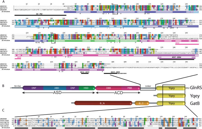
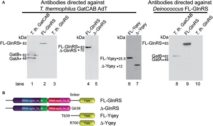
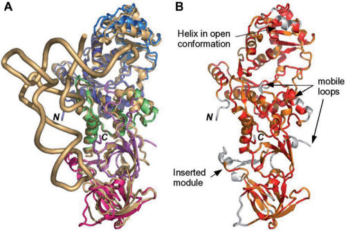
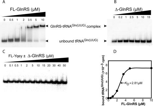
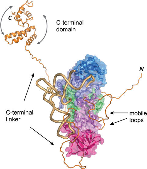
Glutaminyl-tRNA synthetase from Deinococcus radiodurans possesses a C-terminal extension of 215 residues appending the anticodon-binding domain. This domain constitutes a paralog of the Yqey protein present in various organisms and part of it is present in the C-terminal end of the GatB subunit of GatCAB, a partner of the indirect pathway of Gln-tRNA(Gln) formation. To analyze the peculiarities of the structure-function relationship of this GlnRS related to the Yqey domain, a structure of the protein was solved from crystals diffracting at 2.3 A and a docking model of the synthetase complexed to tRNA(Gln) constructed. The comparison of the modeled complex with the structure of the E. coli complex reveals that all residues of E. coli GlnRS contacting tRNA(Gln) are conserved in D. radiodurans GlnRS, leaving the functional role of the Yqey domain puzzling. Kinetic investigations and tRNA-binding experiments of full length and Yqey-truncated GlnRSs reveal that the Yqey domain is involved in tRNA(Gln) recognition. They demonstrate that Yqey plays the role of an affinity-enhancer of GlnRS for tRNA(Gln) acting only in cis. However, the presence of Yqey in free state in organisms lacking GlnRS, suggests that this domain may exert additional cellular functions.
Figures
Similar articles
-
Crystallization and preliminary X-ray characterization of the atypical glutaminyl-tRNA synthetase from Deinococcus radiodurans.Acta Crystallogr D Biol Crystallogr. 2004 Dec;60(Pt 12 Pt 2):2361-3. doi: 10.1107/s0907444904026691. Acta Crystallogr D Biol Crystallogr. 2004. PMID: 15614972
-
Connecting anticodon recognition with the active site of Escherichia coli glutaminyl-tRNA synthetase.J Mol Biol. 1994 Jul 8;240(2):111-8. doi: 10.1006/jmbi.1994.1425. J Mol Biol. 1994. PMID: 8027995
-
Influence of transfer RNA tertiary structure on aminoacylation efficiency by glutaminyl and cysteinyl-tRNA synthetases.J Mol Biol. 2000 Jun 2;299(2):431-46. doi: 10.1006/jmbi.2000.3749. J Mol Biol. 2000. PMID: 10860750
-
Substrate selection by aminoacyl-tRNA synthetases.Nucleic Acids Symp Ser. 1995;(33):40-2. Nucleic Acids Symp Ser. 1995. PMID: 8643392 Review.
-
Selectivity and specificity in the recognition of tRNA by E coli glutaminyl-tRNA synthetase.Biochimie. 1993;75(12):1083-90. doi: 10.1016/0300-9084(93)90007-f. Biochimie. 1993. PMID: 8199243 Review.
Cited by
-
On the evolution of the tRNA-dependent amidotransferases, GatCAB and GatDE.J Mol Biol. 2008 Mar 28;377(3):831-44. doi: 10.1016/j.jmb.2008.01.016. Epub 2008 Jan 16. J Mol Biol. 2008. PMID: 18279892 Free PMC article.
-
Evolutionary insights about bacterial GlxRS from whole genome analyses: is GluRS2 a chimera?BMC Evol Biol. 2014 Feb 12;14:26. doi: 10.1186/1471-2148-14-26. BMC Evol Biol. 2014. PMID: 24521160 Free PMC article.
-
Combining multi-mutant and modular thermodynamic cycles to measure energetic coupling networks in enzyme catalysis.Struct Dyn. 2017 Jan 26;4(3):032101. doi: 10.1063/1.4974218. eCollection 2017 May. Struct Dyn. 2017. PMID: 28191480 Free PMC article.
-
A Ribosomal Protein Homolog Governs Gene Expression and Virulence in a Bacterial Pathogen.J Bacteriol. 2022 Oct 18;204(10):e0026822. doi: 10.1128/jb.00268-22. Epub 2022 Sep 19. J Bacteriol. 2022. PMID: 36121290 Free PMC article.
-
Crystal structure of a transfer-ribonucleoprotein particle that promotes asparagine formation.EMBO J. 2010 Sep 15;29(18):3118-29. doi: 10.1038/emboj.2010.192. Epub 2010 Aug 17. EMBO J. 2010. PMID: 20717102 Free PMC article.
References
-
- Schimmel P, Söll D. Aminoacyl-tRNA synthetases: general features and recognition of transfer RNAs. Annu. Rev. Biochem. 1979;48:601–648. - PubMed
-
- Ibba M, Becker HD, Stathopoulos C, Tumbula DL, Söll D. The adaptor hypothesis revisited. Trends Biochem. Sci. 2000;25:311–316. - PubMed
-
- Tumbula DL, Becker HD, Chang W-Z, Söll D. Domain-specific recruitment of amide amino acids for protein synthesis. Nature. 2000;407:106–110. - PubMed
Publication types
MeSH terms
Substances
LinkOut - more resources
Full Text Sources
Molecular Biology Databases
