. 2007 Mar 1;120(Pt 5):815-25.
doi: 10.1242/jcs.03389. Epub 2007 Feb 6.

Wound-induced ATP release and EGF receptor activation in epithelial cells

Affiliations

Wound-induced ATP release and EGF receptor activation in epithelial cells

Jia Yin et al. J Cell Sci. .

Abstract

We have shown previously that wounding of human corneal epithelial (HCE) cells resulted in epidermal growth factor receptor (EGFR) transactivation through ectodomain shedding of heparin-binding EGF-like growth factor (HB-EGF). However, the initial signal to trigger these signaling events in response to cell injury remains elusive. In the present study, we investigated the role of ATP released from the injured cells in EGFR transactivation in HCE cells as well as in BEAS 2B cells, a bronchial epithelial cell line. Wounding of epithelial monolayer resulted in the release of ATP into the culture medium. The wound-induced rapid activation of phosphatidylinositol-3-kinase (PI3K) and extracellular signal-regulated kinase (ERK) pathways in HCE cells was attenuated by eliminating extracellular ATP, ADP and adenosine. The nonhydrolyzable ATP analog ATP-gamma-S induced rapid and sustained EGFR activation that depended on HB-EGF shedding and ADAM (a disintegrin and metalloproteinase). Targeting pathways leading to HB-EGF shedding and EGFR activation attenuated ATP-gamma-S-enhanced closure of small scratch wounds. The purinoceptor antagonist reactive blue 2 decreased wound closure and attenuated ATP-gamma-S induced HB-EGF shedding. Taken together, our data suggest that ATP, released upon epithelial injury, acts as an early signal to trigger cell responses including an increase in HB-EGF shedding, subsequent EGFR transactivation and its downstream signaling, resulting in wound healing.


Figures
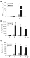
Fig. 1
Fig. 1
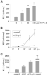
Wounding increases ATP concentration in culture medium of corneal and airway epithelial cells. (A–C) THCE cells (A,B) or BEAS 2B cells (C) were seeded in 60-mm dishes and growth-factor-or serum-starved overnight. Cells were extensively wounded and ATP concentration in the culture medium was measured as described in Materials and Methods. Results are expressed as ATP concentration in μM (A) or relative light units (RLU) (B,C) and data are given as the mean ± s.e.m. (n=3). *P<0.05 and **P<0.01, significant increase of ATP concentration in culture medium after wounding compared with the control; b.w., before wounding, p.w., post wounding.
Fig. 2
Fig. 2
Wounding-induced but not EGFR-ligand-induced AKT and ERK phosphorylation is ATP-dependent. (A) Growth factor-starved THCE cells were pretreated with a combination of apyrase and adenosine deaminase (5 units/ml each) for 30 minutes, and were then either extensively injured or stimulated with HB-EGF (50 ng/ml) as a positive control. After removing debris, cells were further incubated in fresh KBM containing the combination of apyrase and adenosine deaminase or HB-EGF for 2 minutes. Cell lysates were subjected to western blotting with antibodies against phosphorylated AKT (P-AKT), phosphorylated ERK 1/2 (P-ERK), AKT and ERK2. (B) Growth factor-starved THCE cells were pretreated with the apyrase and adenosine deaminase mix as in A and wounded with a 0.1–10 μl pipette tip. Cells were then fixed 15 minutes post wounding and incubated with antibody against PERK, followed by visualization with FITC-conjugated secondary antibody. Corresponding nuclear staining was performed with DAPI. Arrows indicate nuclear staining of P-ERK at the wound edge (arrowhead).
Fig. 3
Fig. 3
ATP-γ-S activates EGFR signaling in corneal and airway epithelial cells. (A–C) Growth-factor-starved THCE cells (A), primary HCE cells (B) or serum-starved BEAS 2B cells (C) were stimulated with 100 μM ATP-γ-S for the indicated times. Cells were then lysed. THCE cell lysates were immunoprecipitated with EGFR antibody, immunoblotted with anti-PY99 antibody (P-EGFR) and re-probed with anti-EGFR antibody (EGFR) to assess the amount of EGFR precipitated. Lysates of THCE, primary HCE and BEAS 2B cells were also subjected to western blotting with antibodies against phosphorylated AKT (P-AKT), phosphorylated ERK1/2 (P-ERK), EGFR Y1068, AKT and ERK2.
Fig. 4
Fig. 4
ATP-γ-S-induced AKT and ERK phosphorylation is Ca2+ and EGFR dependent. Growth-factor-starved THCE cells were pretreated with either 50 μM Ca2+ chelator BAPTA-AM (A) or 1 μM EGFR inhibitor AG 1478 (B) for 1 hour and then stimulated with 100 μM ATP-γ-S for 30 minutes. Cell lysates were subjected to western blotting with antibodies against phosphorylated AKT (P-AKT), phosphorylated ERK1/2 (P-ERK), AKT and ERK2.
Fig. 5
Fig. 5
ATP-γ-S induces HB-EGF release in THCE cells expressing HB-EGF-AP. (A) Cells were cultured in 6-well dishes and treated with different concentrations of ATP-γ-S (1–100 μM) for 20 minutes. AP released into the collected medium was measured as described in Materials and Methods and expressed as relative light units (RLU). (B) Cells were stimulated with 100 μM ATP-γ-S for indicated times post stimulation (p.s.); AP activity was measured and expressed as in A. (C) Cells were stimulated with 100 μM ADP, 100 μM ATP-γ-S, or were extensively wounded; AP activity was measured and expressed as in A. Each RLU represents the mean ± s.e.m. (n=3). *P<0.05 and **P<0.01, significant increase in AP released into the culture medium after ATP-γ-S treatment compared with the control.
Fig. 6
Fig. 6
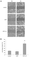
ATP-γ-S, but not ADP, enhances epithelial wound closure. (A,B) Growth-factor-starved THCE cells were wounded with a single tooth of a 48-well sharkstooth comb for DNA sequencing gels. Cells were allowed to heal in KBM containing 100 μM ADP or 100 μM ATP-γ-S. Wound closure was photographed immediately after wounding (0 h) or 6 hours post wounding (6 h). Micrographs (A) represent one of three samples performed each time. Statistical analysis (B) indicates healing extent. Values are expressed as the mean ± s.e.m. (n=3), **P<0.01.
Fig. 7
Fig. 7
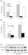
ATP-γ-S-induced HB-EGF shedding and EGFR activation are sensitive to ADAM inhibition. (A,B) Cells expressing HB-EGF-AP were pretreated with (A) 50 μM GM6001 (GM) or its inactive analog (GM neg) or (B) 4 μM GW280264X (GW) for 1 hour and stimulated with 100 μM ATP-γ-S. AP activity was measured and expressed as in Fig. 5. **P<0.01, significant decrease in AP release by the inhibitors in the control and ATP-γ-S-treated cells. NT, no inhibitor treatment. (C) Growth-factor-starved THCE cells were pretreated with 10 μg/ml CRM197 (CRM), 4 μM GW280264X, 50 μM GM6001 or 50 μM GM6001 inactive analog for 1 hour and then stimulated with 100 μM ATP-γ-S for 10 minutes. Cells were then lysed and lysates were subjected to EGFR immunoprecipitation as described in Fig. 3A and phosphorylation determination.
Fig. 8
Fig. 8
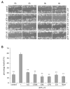
ATP-γ-S enhances epithelial wound closure in an EGFR-, HB-EGF-, ADAM10-, ADAM17-, ERK- and PI3K-dependent manner. Growth-factor-starved THCE cells were pretreated with the following inhibitors 1 μM AG 1478 (AG), 10 μg/ml CRM197 (CRM), 50 μM GM6001 (GM), 4 μM GW280264X (GW), 10 μM U0126 (U0) or 100 nM wortmannin (Wort) for 1 hour, wounded with a single tooth of a 48-well sharkstooth comb for DNA sequencing gels and allowed to heal in KBM containing 100 μM ATP-γ-S in the presence of inhibitors. Wound closure was photographed immediately after wounding (0 h) or 6 hours p.w. (6 h). Micrographs in A represent one of three samples performed each time. Bar graph in B gives the statistical analysis of healing extent. Values are the mean ± s.e.m. (n=3), **P<0.01.
Fig. 9
Fig. 9
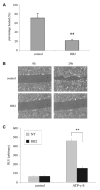
The P2 antagonist RB2 attenuates epithelial wound closure and HB-EGF shedding. (A,B) Growth factor-starved THCE cells were pretreated with 100 μM RB2 for 1 hour and then wounded with a 0.1–10 μl pipette tip. Cells were allowed to heal in KBM with (RB2) or without (control) RB2 for 24 hours. Cell migration was monitored by photographing the wound immediately (0 h) or 24 hours post wounding (24 h). Micrographs in A represent one of three samples performed each time. Statistical analysis of healing extent is shown in B. Values are the mean ± s.e.m. (n=3), **P<0.01. (C) Cells expressing HB-EGF-AP were pretreated with (RB2) or without (NT) 10 μm RB2 and stimulated with 100 μM ATP-γ-S. AP activity was measured and expressed as RLU. **P<0.01, significant inhibition in AP release by RB2 in the control or ATP-γ-S-treated cells.
Fig. 10
Fig. 10
Proposed extracellular ATP involvement in corneal epithelial wound healing that summarizes the pathway from ATP release caused by cell injury to wound closure, including P2Y activation, intracellular Ca2+ release and EGFR signaling. Wounding results in the release of ATP from damaged cells, and the released ATP –through its P2 receptors – triggers intracellular Ca2+ waves leading to activation of ADAM protein(s) in a neighboring cell that is not injured (△). ADAM protein(s) cleaves pro-HB-EGF at the cell surface and HB-EGF in an autocrine and/or paracrine fashion binds EGFR, which transduces the signals into the intracellular signaling network. After EGFR activation, HCE cell migration, proliferation and wound healing are induced via PI3K, ERK and other intracellular signaling pathways.

Similar articles

Cited by

References

    1. Agteresch HJ, Dagnelie PC, van der Gaast A, Stijnen T, Wilson JH. Randomized clinical trial of adenosine 5′-triphosphate in patients with advanced non-small-cell lung cancer. J Natl Cancer Inst. 2000;92:321–328. - PubMed
    1. Agteresch HJ, Rietveld T, Kerkhofs LG, van den Berg JW, Wilson JH, Dagnelie PC. Beneficial effects of adenosine triphosphate on nutritional status in advanced lung cancer patients: a randomized clinical trial. J Clin Oncol. 2002;20:371–378. - PubMed
    1. Ahmad S, Ahmad A, McConville G, Schneider BK, Allen CB, Manzer R, Mason RJ, White CW. Lung epithelial cells release ATP during ozone exposure: signaling for cell survival. Free Radic Biol Med. 2005;39:213–226. - PubMed
    1. Araki-Sasaki K, Ohashi Y, Sasabe T, Hayashi K, Watanabe H, Tano Y, Handa H. An SV40-immortalized human corneal epithelial cell line and its characterization. Invest Ophthalmol Vis Sci. 1995;36:614–621. - PubMed
    1. Blobel CP. Functional and biochemical characterization of ADAMs and their predicted role in protein ectodomain shedding. Inflamm Res. 2002;51:83–84. - PubMed

Publication types

MeSH terms

Substances