. 2007 May 15;109(10):4320-7.
doi: 10.1182/blood-2006-11-053769. Epub 2007 Feb 6.

DC-HIL is a negative regulator of T lymphocyte activation

Affiliations

DC-HIL is a negative regulator of T lymphocyte activation

Jin-Sung Chung et al. Blood. .

Abstract

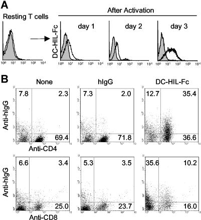
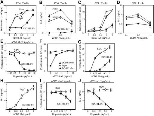
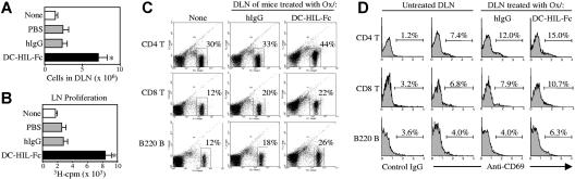
T-cell activation is the net product of competing positive and negative signals transduced by regulatory molecules on antigen-presenting cells (APCs) binding to corresponding ligands on T cells. Having previously identified DC-HIL as a receptor expressed by APCs that contains an extracellular immunoglobulin (Ig)-like domain, we postulated that it plays a role in T-cell activation. To probe this function, we created soluble recombinant DC-HIL, which we observed to bind activated (but not resting) T cells, indicating that expression of the putative ligand on T cells is induced by activation. Binding of DC-HIL to naive T cells attenuated these cells' primary response to anti-CD3 antibody, curtailing IL-2 production, and preventing entry into the cell cycle. DC-HIL also inhibited reactivation of T cells previously activated by APCs (secondary response). By contrast, addition of soluble DC-HIL to either allogeneic or ovalbumin-specific lymphocyte reactions augmented T-cell proliferation, and its injection into mice during the elicitation (but not sensitization) phase of contact hypersensitivity exacerbated ear-swelling responses. Mutant analyses showed the inhibitory function of DC-HIL to reside in its extracellular Ig-like domain. We conclude that endogenous DC-HIL is a negative regulator of T lymphocyte activation, and that this native inhibitory function can be blocked by exogenous DC-HIL, leading to enhanced immune responses.


Figures

Figure 1
Figure 1
DC-HIL-Fc binds to activated (but not resting) T cells. (A) Purified splenic CD4+ T cells were cultured with concanavalin A (10 μg/mL) for 3 days. After blocking the binding activity of Fc receptors on cells with a Fc blocker, treated T cells were stained with DC-HIL-Fc/ FITC-anti-human IgG Ab (open histograms), or corresponding control Abs (gray histograms). Binding of DC-HIL-Fc to T cells was analyzed by FACS. (B) Purified CD3+ T cells were treated with immobilized anti-CD3 Ab (1 μg/mL) for 3 days and then stained with FITC-labeled anti-CD4 or anti-CD8 Ab and with DC-HIL-Fc or hIgG/PE-anti-human IgG. Numbers in each quadrant represent percentages of the total cell population. Data shown are representative of 3 independent experiments.
Figure 2
Figure 2
Immobilized DC-HIL-Fc inhibits T-cell activation triggered by anti-CD3 Ab. CD4+ (A-B) or CD8+ (C-D) T cells (2 × 10⁵ each) purified from BALB/c spleens were cultured for 48 hours in microculture wells precoated with increasing doses of anti-CD3 Ab and with a constant dose (10 μg/mL) of DC-HIL-Fc (●), control hIgG (○), or neither (none; ▵). After pulsing with 3H-thymidine, cells and culture supernatant were harvested. 3H-thymidine incorporation into cells (A,C) and IL-2 production (B,D) were determined, and values (cpm or ng/mL) plotted at a logarithmic scale, respectively. (E) Titration of inhibitory function of DC-HIL-Fc. CD4+ T cells were cultured for 48 hours in microculture wells precoated with a constant dose (0.3 μg/mL) of anti-CD3 Ab and increasing doses of DC-HIL-Fc or control hIgG. (F) CD28 costimulation rescues DC-HIL-Fc–induced inhibition of CD4+ T-cell activation. Purified CD4+ T cells were cultured in wells precoated with anti-CD3 Ab (0.3 μg/mL), hIgG or DC-HIL-Fc (5 μg/mL), and anti-CD28 mAb (increasing doses). (G-H) Previously activated T cells were prepared from Tac-TgN(DO11.10)-Rag2tm1mice and reactivated by immobilized anti-CD3 Ab (varying doses) and DC-HIL-Fc or hIgG (constant dose). (I-J) Inhibitory function of DC-HIL-Fc was titrated against reactivation of previously activated T cells by anti-CD3 Ab (1 μg/mL). Proliferation (G,I) and IL-2 production (H,J) were measured. Results are expressed as mean values ± SDs. Data shown are representative of 6 (A-B), 3 (G-J), and 2 (C-F) experiments, respectively.
Figure 3
Figure 3
Cell-cycle analyses of CD4+ T cells treated with DC-HIL-Fc. (A) T cells (6 × 10⁶) were labeled with CFSE (1 μM) and then cultured in microwells precoated with anti-CD3 Ab (0.3 μg/mL) plus hIgG or DC-HIL-Fc (5 μg/mL). At the indicated time points, cells were harvested and analyzed by FACS for fluorescence intensity. The frequency (%) of divided cells is shown in histograms. (B) T cells from 48-hour culture similarly treated were analyzed for incorporation of BrdU (using FITC–anti-BrdU Ab) and total DNA content (stained with 7-AAD) by FACS; data shown as dot plots of BrdU versus 7-AAD. Data shown are representative of 3 (A) and 2 (B) independent experiments.
Figure 4
Figure 4
Soluble DC-HIL-Fc enhances responses of CD4+ T cells by APCs. Effects of soluble DC-HIL-Fc on T-cell activation were examined in MLR (A-B), anti-CD3 response (C), or in OVA-specific antigen presentation (D-E). MLR: C57BL/6 spleen cells (5 × 10⁴) were γ irradiated and mixed with CD4+ T cells (2 × 10⁵) purified from BALB/c mouse spleens. (A) Increasing doses of hIgG or DC-HIL-Fc were added to the MLR culture and incubated for 2 days prior to 3H-thymidine pulsing. Proliferative response of T cells was assayed by incorporation of 3H-thymidine. (B) MLR was incubated in the absence/presence of hIgG or DC-HIL-Fc (20 μg/mL) for 1, 2, or 3 days before pulsing. (C) Soluble (Sol) DC-HIL-Fc does not inhibit T-cell activation triggered by immobilized (Im) anti-CD3 Ab. CD4+ T cells were cultured in microwells precoated with anti-CD3 Ab (0.3 μg/mL) and 5 μg/mL of DC-HIL-Fc. In some wells, soluble hIgG or DC-HIL-Fc in increasing doses was added to culture in wells coated with the same amount of anti-CD3 Ab. T-cell activation was expressed as proliferative capacity. (D-E) OVA-specific response: CD4+ T cells purified from the spleens of BALB/cTac-TgN(DO11.10)-Rag2tm1 mice were cocultured without (No) or with BM-DCs (from BALB/c mice) previously pulsed with OVA peptide. T-cell activation was assayed by IL-2 production (D) and by FACS for frequency of CD69+/CD4+ T cells (E). Control staining was performed with FITC-rat IgG (rIgG) and PE-hamster IgG (haIgG). (F) siRNA-mediated knockdown of DC-HIL. At 1 day after transfection of DCs with control (Ctrl; shuffled) siRNA or DC-HIL–targeted siRNA, cells were harvested and assayed by immunoblotting for protein expression of DC-HIL or β-actin. (G) Increasing numbers of transfected DCs were pulsed with OVA peptide and cocultured with a constant number of OVA-specific CD4+ T cells. Activation was measured by IL-2 production. (H) At 2 days after coculturing, frequency of CD69+ in the CD4+ T cells was determined by FACS. *Statistical significance (P < .001) compared with T-cell responses treated with hIgG control. Data shown are representative of at least 3 independent experiments.
Figure 5
Figure 5
Soluble DC-HIL-Fc enhances elicitation of Ox-induced contact hypersensitivity in mice. Sensitization of BALB/c mice (n = 5) with Ox for CH (A-B): on day 0, mice were painted with 2% Ox on abdominal skin (Senst). On day 6, CH was elicited in sensitized mice by painting 1% Ox or solvent control to right and left ears, respectively (Challenge). CH was assessed daily through day 9 or 12 by measuring ear thickness (▴). Mice were injected intraperitoneally with PBS, hIgG, or DC-HIL-Fc (10 mg/kg each) on days −1, 1, and 3 (before and after sensitization) (A) or on days 5, 7, and 9 (before and after challenge) (B). Daily change in ear thickness was plotted for each panel during sensitization (A) or elicitation (B). *P < .003; **P < .05 compared with ear thickness of mice treated with hIgG. (C) Ear skin was excised from mice treated without (None) or with Ox and Fc protein (2 days after elicitation) and examined histologically (10×/10 objective lens). Data shown (A-B) are representative of 4 independent experiments.
Figure 6
Figure 6
Ox/DC-HIL-Fc–treated LN cells display hyperactivation phenotypes. In an independent experiment, draining LN (DLN) cells prepared from BALB/c mice treated similarly (as in Figure 5) were examined. (A) DLN cells were counted. (B) Spontaneous activation was measured by 3H-thymidine incorporation of DLN cells (4 × 10⁵/well) cultured for 3 days without stimuli. (C-D) frequency of leukocytes: DLN cells were stained with FITC-Ab against CD4, CD8, or B220 alone (C) or doubly stained with PE–anti-CD69 (D), and then analyzed by FACS. CD69 expression (D) is shown in LN cells stained positively with the surface marker Ab. Results (A and B) are shown as mean values ± SDs; *P < .001 compared with LN responses treated with hIgG control. Data shown are representative of 3 independent experiments.
Figure 7
Figure 7
Mutant analyses of DC-HIL-Fc function. (A) Protein structures of DC-HIL-Fc wild-type (WT) and mutants are represented schematically. Extracelluar domains (ECDs) of mutants, RAA (replacement of RGD sequence with RAA; ▴) and deletion mutants lacking PRR (amino acid [aa] 301-334) and PKD230-355 were linked to a Fc portion of hIgG and produced in COS-1 cells. (B) After purifying mutant DC-HIL-Fc proteins, a small aliquot (2 μg/lane) was run on SDS-PAGE and then stained with Coomassie Blue to visualize protein bands. (C) T-cell activation. Highly purified DC-HIL-Fc WT or mutants (5 μg/mL each) and anti-CD3 Ab (increasing doses) were coated on microwells for culture with CD4+ T cells for 2 days and pulsed with 3H-thymidine for 20 hours. Results are shown as mean values ± SDs. (D) Binding of DC-HIL mutants to T cells. Activated CD4+ T cells were incubated with WT and mutants of DC-HIL (10 μg/mL) and analyzed for binding by FACS. Histograms of T cells stained with hIgG (filled) and a mutant (open) are overlaid. Data (C-D) shown are representative of 3 experiments.

Similar articles

Cited by

References

    1. Chambers CA, Allison JP. Co-stimulation in T cell responses. Curr Opin Immunol. 1997;9:396–404. - PubMed
    1. Acuto O, Michel F. CD28-mediated co-stimulation: a quantitative support for TCR signalling. Nat Rev Immunol. 2003;3:939–951. - PubMed
    1. Walunas TL, Lenschow DJ, Bakker CY, et al. CTLA-4 can function as a negative regulator of T cell activation. Immunity. 1994;1:405–413. - PubMed
    1. Krummel MF, Allison JP. CD28 and CTLA-4 have opposing effects on the response of T cells to stimulation. J Exp Med. 1995;182:459–465. - PMC - PubMed
    1. Tivol EA, Borriello F, Schweitzer AN, et al. Loss of CTLA-4 leads to massive lymphoproliferation and fatal multiorgan tissue destruction, revealing a critical negative regulatory role of CTLA-4. Immunity. 1995;3:541–547. - PubMed

Publication types

MeSH terms