. 2007 May 15;109(10):4392-8.
doi: 10.1182/blood-2006-03-012575. Epub 2007 Feb 6.

The p21Waf1 pathway is involved in blocking leukemogenesis by the t(8;21) fusion protein AML1-ETO

Affiliations

The p21Waf1 pathway is involved in blocking leukemogenesis by the t(8;21) fusion protein AML1-ETO

Luke F Peterson et al. Blood. .

Abstract

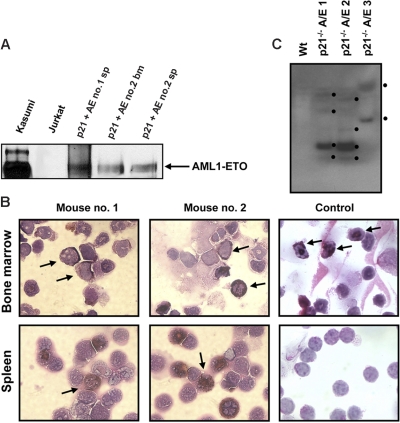
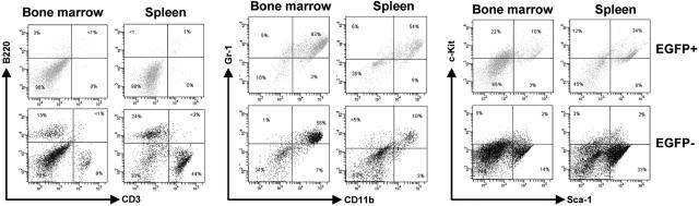
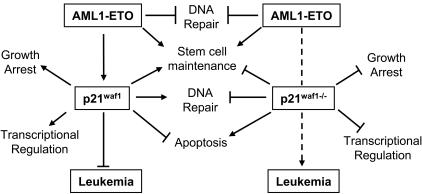
The 8;21 translocation is a major contributor to acute myeloid leukemia (AML) of the M2 classification occurring in approximately 40% of these cases. Multiple mouse models using this fusion protein demonstrate that AML1-ETO requires secondary mutagenic events to promote leukemogenesis. Here, we show that the negative cell cycle regulator p21(WAF1) gene is up-regulated by AML1-ETO at the protein, RNA, and promoter levels. Retroviral transduction and hematopoietic cell transplantation experiments with p21(WAF1)-deficient cells show that AML1-ETO is able to promote leukemogenesis in the absence of p21(WAF1). Thus, loss of p21(WAF1) facilitates AML1-ETO-induced leukemogenesis, suggesting that mutagenic events in the p21(WAF1) pathway to bypass the growth inhibitory effect from AML1-ETO-induced p21(WAF1) expression can be a significant factor in AML1-ETO-associated acute myeloid leukemia.


Figures

Figure 1
Figure 1
AML1-ETO regulates the cell cycle inhibitor p21WAF1. (A) K562 cells infected with MigR1 and Mig-A/E were sorted by flow cytometry 2 days after infection for EGFP+ cells and cultured overnight at 2 × 10⁵ cells/mL and harvested for cell cycle analysis by propidium iodide staining by flow cytometry. Percentages of G0/G1 and cells in S/G2/M phase of cells are presented from 3 individual infection experiments. (B) Following infection and flow cytometry sorting were analyzed by Western transfer with 20 μg protein the blots were probed with anti-HA or anti-p21waf1 to analyze their expression. Ponceau staining of the blot depicts loading. (C) K562 cells were infected, sorted, and cultured as described and RNA was prepared. RNA (10 μg) was analyzed by Northern blot with the full-length p21WAF1 cDNA. Equal loading is depicted by the 18S ribosomal RNA. (D) Schematic diagram of the 2.3-kb promoter region of the p21WAF1 gene cloned into the pGL2 reporter vector; ■, AML1 DNA-binding sites; ▨, putative AML1-binding site (TGTGGG). (E) K562 cells were transfected with 10 μg p21-pGL2, with either 2 μg empty vector or those expressing HA-AML1-ETO, HA-AML1-ETO-R174Q, AML1-ETO, or AML1-ETO-L148D brought to a total of 20μg with 5.6 μg herring sperm DNA, 2 μg of an expression plasmid for CBFβ and 400 ng pRL-null as internal control for transfection, and cultured for 16 to 23 hours. Experiments depict the average and SD of 5 individual experiments with 2 different batches of DNA. (F) ChIP assay for association of AML1 and AML1-ETO to the distal and proximal regions of the p21WAF1 promoter. Cells were transfected with 10 μg p21pGL2 or Δ1.9p21pGL2 with either 2 μg pCDNA6, pCDNA6-HA-AML1, or pCDNA6-HA-AML1-ETO and following ChIP the isolated DNA fragments were subjected to PCR with specific primers that amplify regions 1 and 2 depicted in panel D, and analyzed by DNA gel electrophoresis.
Figure 2
Figure 2
AML1-ETO promotes leukemia in p21Waf1-deficient mice. (A) Kaplan-Meier survival plot of mice receiving transplants of MigR1- or Mig-A/E–infected p21Waf1 cells. Mice were monitored for leukemia development 3 weeks after transplantation. (B) Hematoxylin and eosin staining of liver and spleen of a control mouse and a leukemic mouse, showing the disruption of the tissue architecture by infiltrating leukemic cells. (C) Accumulation of blast cells derived from AML1-ETO–expressing p21Waf1-deficient cells in hematopoietic compartments. Cells isolated from spleen and bone marrow from a recipient mouse of transplanted p21WAF1-deficient cells expressing AML1-ETO and a control were cytospun and stained with Wright-Giemsa for the analysis of immature blasts indicated by the arrows. Microscopy was performed with a Leica DMLB microscope (Leica Microsystems, Wetzlar, Germany) using an HC PLAN 10×/22 eyepiece and 10×/0.25 or 20×/0.4 objective lenses, or a 100×/1.30 oil-immersion objective lens (Richard Allan Scientific, Kalamazoo, MI). Pictures were captured using Spot software from Diagnostic Instruments (Sterling Heights, MI).
Figure 3
Figure 3
Expression of AML1-ETO, MPO, and clonality in leukemic cells. (A) Western blot analysis of 2 independent mice spleen cell samples and one bone marrow cell sample showing the expression of AML1-ETO. Kasumi cells serve as a positive control. (B) Bone marrow and spleen cells from recipient mice of p21Waf1-deficient cells expressing AML1-ETO and a control mouse were cytospun and stained for MPO and counterstained with Wright-Giemsa for the analysis of immature blasts. The arrows indicate MPO+ immature blasts in the leukemic mice and in the control sample MPO+ neutrophils in the bone marrow. Image acquisition was performed as described for Figure 2, using a 100×/1.30 oil-immersion objective lens. (C) gDNA was isolated from 3 leukemic p21Waf1−/− mice expressing AML1-ETO. The DNA was digested with BamHI and a Southern blot prepared. The blot was hybridized with the ETO probe 5′ of the BamHI site in ETO, washed, and exposed to film. The black dots indicate the integration signals.
Figure 4
Figure 4
Flow cytometric analysis of AML1-ETO–expressing p21WAF1-deficient cells. Bone marrow and spleen cells were collected from leukemic mice transplanted with p21Waf1−/−-expressing AML1-ETO cells and analyzed for the expression of CD3/B220, Gr-1/CD11b, and c-kit/Sca-1. Depicted are the EGFP+ and EGFP cells within a particular mouse.
Figure 5
Figure 5
Model of AML1-ETO leukemogenesis through the p21WAF1 pathway. Here we propose a model whereby the bypass of the p21WAF1-regulated pathway(s) cooperates with AML1-ETO in promoting leukemia development. Specifically, in the presence of p21WAF1 AML1-ETO ability to promote leukemia is limited by p21WAF1 role in growth regulation, genome stability, regulating transcription networks, and in primary cells' apoptosis evasion. However, both p21WAF1 and AML1-ETO regulate stem cell maintenance and expansion. The loss of p21WAF1 role in growth regulation, DNA repair, and regulation of transcription establishes an advantageous situation for leukemia development.

References

    1. Meyers S, Downing JR, Hiebert SW. Identification of AML-1 and the (8;21) translocation protein (AML-1/ETO) as sequence-specific DNA-binding proteins: the runt homology domain is required for DNA binding and protein-protein interactions. Mol Cell Biol. 1993;13:6336–6345. - PMC - PubMed
    1. Licht JD. AML1 and the AML1-ETO fusion protein in the pathogenesis of t(8;21) AML. Oncogene. 2001;20:5660–5679. - PubMed
    1. Look AT. Fusion genes and their hybrid proteins in human leukemias and lymphomas. Proc Assoc Am Physicians. 1995;107:175–180. - PubMed
    1. Mikhail FM, Sinha KK, Saunthararajah Y, Nucifora G. Normal and transforming functions of RUNX1: A perspective. J Cell Physiol. 2006;207:582–593. - PubMed
    1. Miyoshi H, Shimizu K, Kozu T, et al. t(8;21) breakpoints on chromosome 21 in acute myeloid leukemia are clustered within a limited region of a single gene, AML1. Proc Natl Acad Sci U S A. 1991;88:10431–10434. - PMC - PubMed

Publication types

MeSH terms

Substances