. 2007 Feb 13;104(7):2301-6.
doi: 10.1073/pnas.0608382104. Epub 2007 Feb 6.

Acute postnatal ablation of Hif-2alpha results in anemia

Affiliations

Acute postnatal ablation of Hif-2alpha results in anemia

Michaela Gruber et al. Proc Natl Acad Sci U S A. .

Abstract

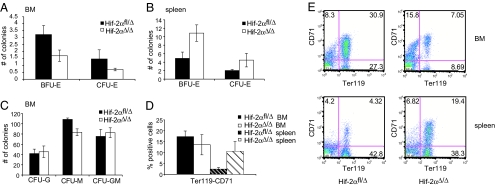
Adaptive transcriptional responses to oxygen deprivation (hypoxia) are mediated by the hypoxia-inducible factors (HIFs), heterodimeric transcription factors composed of two basic helix-loop-helix-PAS family proteins. The transcriptional activity of HIF is determined by the hypoxic stabilization of the HIF-alpha proteins. HIF-1alpha and HIF-2alpha exhibit high sequence homology but have different mRNA expression patterns; HIF-1alpha is expressed ubiquitously whereas HIF-2alpha expression is more restricted to certain tissues, e.g., the endothelium, lung, brain, and neural crest derivatives. Germ-line deletion of either HIF subunit is embryonic lethal with unique features suggesting important roles for both HIF-alpha isoforms. Global deletion of Hif-2alpha results in distinct phenotypes depending on the mouse strain used for the mutation, clearly demonstrating an important role for HIF-2alpha in mouse development. The function of HIF-2alpha in adult life, however, remains incompletely understood. In this study, we describe the generation of a conditional murine Hif-2alpha allele and the effect of its acute postnatal ablation. Under very stringent conditions, we ablate Hif-2alpha after birth and compare the effect of acute global deletion of Hif-2alpha and Hif-1alpha. Our results demonstrate that HIF-2alpha plays a critical role in adult erythropoiesis, with acute deletion leading to anemia. Furthermore, although HIF-1alpha was first purified and cloned based on its affinity for the human erythropoietin (EPO) 3' enhancer hypoxia response element (HRE) and regulates Epo expression during mouse embryogenesis, HIF-2alpha is the critical alpha isoform regulating Epo under physiologic and stress conditions in adults.


Conflict of interest statement

The authors declare no conflict of interest.

Figures

Fig. 1.
Fig. 1.
Generation of a conditional Hif-2α allele. (A) Schematic of the targeting vector and recombination steps. (B) PCR genotyping of mice with Cre activated as young pups. Lanes 1 –3 show Hif-2αfl/fl mice without Cre expression. Lane 4* shows a Hif-2αfl animal without Cre expression. Samples in lanes 5–7 represent DNA from mice which harbor the Cre transgene and show loss of Hif-2α. (C) Southern blot analysis of DNA isolated from the kidney (lanes 1, 4, 7), the liver (lanes 2, 5, 8), and the lung (lanes 3, 6, 9) of Cre negative and positive animals. (D) RT-PCR analysis of cDNA from kidneys and livers of six different animals. Lanes 1 and 7 show floxed mRNA transcripts still containing exon 2 (650 bp). Lanes 2*, 3*, 8*, and 9* express both the floxed and deleted exon 2 mRNA, and a background band. Depletion of Hif-2α results in total loss of exon 2 as displayed in lanes 4–6, and 10–12 (450 bp). (E) β-Galactosidase expression after Cre activation in ROSA26 reporter mice.
Fig. 2.
Fig. 2.
Hif-2αΔ/Δ mice are anemic. (AC) Total red blood cell (RBC) numbers (A), hemoglobin concentration (B) and hematocrit levels (C) are reduced 25%, 32%, and 30%, respectively, in mice with high efficiency of Hif-2α exon 2 deletion. Medium deletion leads to a drop of only 8% (RBCs), 14% (hemoglobin), and 10% (hematocrit levels), and low efficiency of Hif-2α exon 2 deletion does not show a decrease compared with Hif-2αfl levels. (D) PCR analysis of DNA isolated from earpunches taken 2 weeks after Cre activation. Lanes 1–7 are negative for Cre and therefore either fl/wt (lanes 2, 6, and 7) or fl/Δ (lanes 1 and 3–5). Lanes 8–16 are positive for the Cre transgene and according to PCR have lost 90–100% of Hif-2α expression (lanes 8–10, and 13) or ≈75% of Hif-2α (lanes 12, 14, and 15), or show very weak loop-out, in which case the animal resembles a Hif-2α heterozygous (lanes 11 and 16). (E) Activation of Cre during the first week after birth results in a uniformly high deletion efficiency and leads to a 32% drop in spun hematocrit value. (F) Mice that exhibit Hif-1α deletion fail to develop anemia. (G) Loop-out efficiency for Hif-1α.
Fig. 3.
Fig. 3.
Erythroid progenitor cells are decreased in Hif-2αΔ/Δ bone marrow but increased in the spleen. (A) Hif-2αΔ/Δ bone marrow progenitor cells form fewer erythroid colonies in methylcellulose than Hif-2αfl cells do (n = 4). (B) In contrast, Hif-2αΔ/Δ splenic progenitor cells have a higher potential to form erythroid colonies than cells from Hif-2αfl spleens (n = 4). (C) There is little difference in the number of nonerythroid colonies formed by Hif-2αfl and Hif-2αΔ/Δ bone marrow cells (n = 4). (D) The percentage of Ter119high/CD71high erythroid progenitor cells is reduced in Hif-2αΔ/Δ bone marrow, but increased in Hif-2αΔ/Δ spleen (n = 3). (E) Representative FACS blots for Hif-2αfl and Hif-2αΔ/Δ bone marrow and spleen stained for Ter119 and CD71.
Fig. 4.
Fig. 4.
Epo expression after stress-induced erythropoiesis depends on HIF-2α. (A) Spleen-to-body weight ration of PH untreated and treated Hif-2αfl and Hif-2αΔ/Δ mice. (B) Serum EPO levels from PH treated Hif-2α fl/fl, fl/Δ, and Δ/Δ mice. (C) Hif-1α fl/fl and Δ/Δ mice do not exhibit any defects in EPO stimulation after PH treatment. (D) Hematocrit values increase in Hif-2αfl and Hif-2αΔ/Δ animals because of EPO treatment and reach normal levels after 2 weeks in Hif-2αΔ/Δ mice.
Fig. 5.
Fig. 5.
In vitro analysis of the ability of HIF-1α and HIF-2α to induce EPO mRNA transcription. (A) siRNA knockdown of HIF-1α and HIF-2α. Western blot for HIF-1α and HIF-2α shows stabilization of the protein under hypoxia (H) and specific loss of the appropriate protein after siRNA transfection. (B) EPO mRNA levels in Hep3B cells. Fold change of mRNA under hypoxia compared with normoxic cells. Nontransfected cells and cells transfected with control siRNA show a 55-fold induction of EPO mRNA under hypoxia. Knocking-down HIF-1α results in a 45-fold increase in EPO mRNA. In contrast, siRNA against HIF-2α resulted in a 5-fold induction and knocking-down both HIFs almost completely ablates EPO mRNA induction under hypoxia. (C) A mutated (“TM”) and therefore stabilized form of HIF-1α and HIF-2α was introduced into Hep3B cells at increasing concentrations, resulting in a strong induction of EPO mRNA for HIF-1α and a less prominent induction for HIF-2α. (D) Western blot displaying the levels of overexpressed protein.

References

    1. Schofield CJ, Ratcliffe PJ. Nat Rev Mol Cell Biol. 2004;5:343–354. - PubMed
    1. Semenza GL. Trends Mol Med. 2001;7:345–350. - PubMed
    1. Giaccia AJ, Simon MC, Johnson R. Genes Dev. 2004;18:2183–2194. - PMC - PubMed
    1. Ivan M, Kondo K, Yang H, Kim W, Valiando J, Ohh M, Salic A, Asara JM, Lane WS, Kaelin WG., Jr Science. 2001;292:464–468. - PubMed
    1. Jaakkola P, Mole DR, Tian YM, Wilson MI, Gielbert J, Gaskell SJ, Kriegsheim A, Hebestreit HF, Mukherji M, et al. Science. 2001;292:468–472. - PubMed

Publication types

Substances