

Wnt signaling interacts with Shh to regulate taste papilla development
- PMID: 17284610
- PMCID: PMC1794217
- DOI: 10.1073/pnas.0607399104
Wnt signaling interacts with Shh to regulate taste papilla development
Abstract
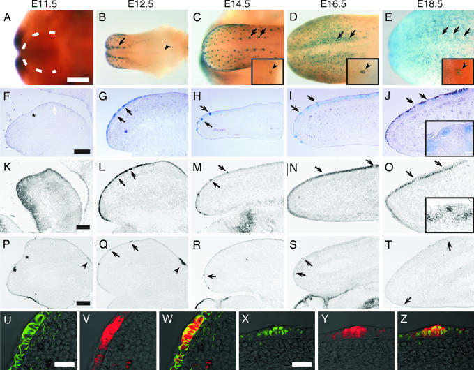
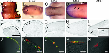
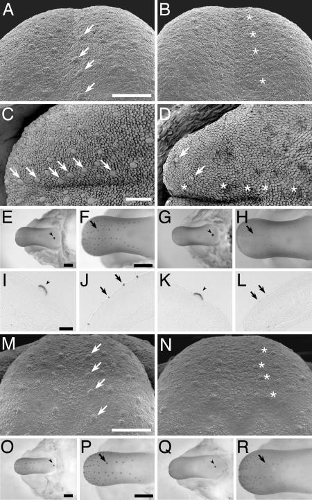
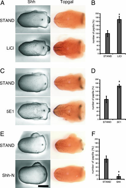
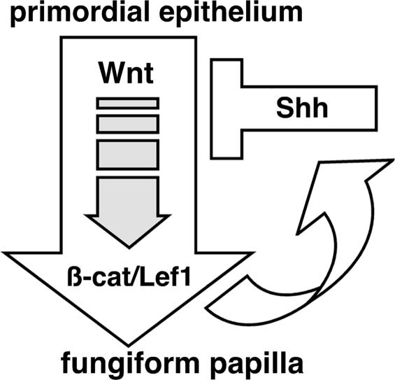
Wnt and Shh signaling pathways are critical for the development and maturation of many epithelial tissues. Both pathways have roles in stem cell maintenance, tissue development, and tumorigenesis. However, linkage between these pathways in mammalian systems had not been well established. Here, we report that Shh expression in fungiform papillae and formation of normal mature fungiform papillae depend on signaling through Wnt and beta-catenin. We observed that during fungiform papilla formation in mice, Shh and components of the Wnt/beta-catenin signaling pathway are expressed together in the developing placode. The elimination of Wnt/beta-catenin signaling in either Lef1 or Wnt10b knockout mice resulted in down-regulation of Shh expression. In addition, the size and number of fungiform papillae were greatly reduced in Lef1 knockout mice. By examining embryonic mouse tongues in culture we determined that activation of Wnt/beta-catenin signaling up-regulates Shh expression. We observed that blocking Shh signaling in cultured tongue explants enhanced papillae formation and was accompanied by an up-regulation of Wnt/beta-catenin signaling, indicating that Shh inhibits the Wnt/beta-catenin pathway. Exogenously added Shh suppressed expression of endogenous Shh and inhibited Wnt/beta-catenin signaling (assessed in TOPGAL mice), further implicating Shh as an inhibitor of the Wnt/beta-catenin pathway. Our observations indicate that Wnt/beta-catenin signaling and interactions between the Wnt and Shh pathways play essential roles in the development of fungiform papillae.
Conflict of interest statement
Conflict of interest statement: R.F.M. has a personal financial interest in the form of stock ownership in Linguagen Corp., receives consulting fees from Linguagen Corp., and is an inventor on patents and patent applications in the area of taste signal transduction that have been licensed to Linguagen Corp. Linguagen Corp. carries out work in the area of taste modification and taste signaling. The work described in the present manuscript would appear to be only peripherally related to the interests of Linguagen Corp.
Figures
Similar articles
-
Gpr177-mediated Wnt Signaling is Required for Fungiform Placode Initiation.J Dent Res. 2014 Jun;93(6):582-8. doi: 10.1177/0022034514531985. Epub 2014 Apr 15. J Dent Res. 2014. PMID: 24736288
-
Sonic hedgehog exerts distinct, stage-specific effects on tongue and taste papilla development.Dev Biol. 2004 Dec 15;276(2):280-300. doi: 10.1016/j.ydbio.2004.07.042. Dev Biol. 2004. PMID: 15581865
-
β-Catenin signaling regulates temporally discrete phases of anterior taste bud development.Development. 2015 Dec 15;142(24):4309-17. doi: 10.1242/dev.121012. Epub 2015 Nov 2. Development. 2015. PMID: 26525674 Free PMC article.
-
Wnt and hedgehog signaling pathways in bone development.J Bone Joint Surg Am. 2008 Feb;90 Suppl 1:19-24. doi: 10.2106/JBJS.G.01174. J Bone Joint Surg Am. 2008. PMID: 18292352 Review.
-
Wnt/beta-catenin signaling in oral tissue development and disease.J Dent Res. 2010 Apr;89(4):318-30. doi: 10.1177/0022034510363373. Epub 2010 Mar 3. J Dent Res. 2010. PMID: 20200414 Free PMC article. Review.
Cited by
-
Lgr5-EGFP marks taste bud stem/progenitor cells in posterior tongue.Stem Cells. 2013 May;31(5):992-1000. doi: 10.1002/stem.1338. Stem Cells. 2013. PMID: 23377989 Free PMC article.
-
Retinoic acid alters the proliferation and survival of the epithelium and mesenchyme and suppresses Wnt/β-catenin signaling in developing cleft palate.Cell Death Dis. 2013 Oct 31;4(10):e898. doi: 10.1038/cddis.2013.424. Cell Death Dis. 2013. PMID: 24176856 Free PMC article.
-
The odontode explosion: the origin of tooth-like structures in vertebrates.Bioessays. 2010 Sep;32(9):808-17. doi: 10.1002/bies.200900151. Bioessays. 2010. PMID: 20730948 Free PMC article. Review.
-
Lrp4 and Wise interplay controls the formation and patterning of mammary and other skin appendage placodes by modulating Wnt signaling.Development. 2013 Feb 1;140(3):583-93. doi: 10.1242/dev.085118. Development. 2013. PMID: 23293290 Free PMC article.
-
[Development and homeostasis of taste buds in mammals].Hua Xi Kou Qiang Yi Xue Za Zhi. 2018 Oct 1;36(5):552-558. doi: 10.7518/hxkq.2018.05.016. Hua Xi Kou Qiang Yi Xue Za Zhi. 2018. PMID: 30465351 Free PMC article. Chinese.
References
-
- Hooper JE, Scott MP. Nat Rev Mol Cell Biol. 2005;6:306–317. - PubMed
-
- Logan CY, Nusse R. Annu Rev Cell Dev Biol. 2004;20:781–810. - PubMed
-
- Gregorieff A, Clevers H. Genes Dev. 2005;19:877–890. - PubMed
-
- Taipale J, Beachy PA. Nature. 2001;411:349–354. - PubMed
-
- McMahon AP, Ingham PW, Tabin CJ. Curr Top Dev Biol. 2003;53:1–114. - PubMed
Publication types
MeSH terms
Substances
Grants and funding
LinkOut - more resources
Full Text Sources
Molecular Biology Databases
