. 2007 Apr;17(4):451-60.
doi: 10.1101/gr.5651507. Epub 2007 Feb 6.

AT-rich repeats associated with chromosome 22q11.2 rearrangement disorders shape human genome architecture on Yq12

Affiliations

AT-rich repeats associated with chromosome 22q11.2 rearrangement disorders shape human genome architecture on Yq12

Melanie Babcock et al. Genome Res. 2007 Apr.

Abstract

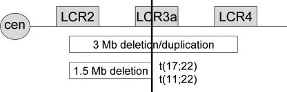
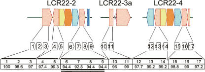
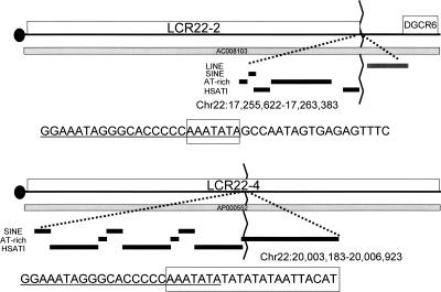
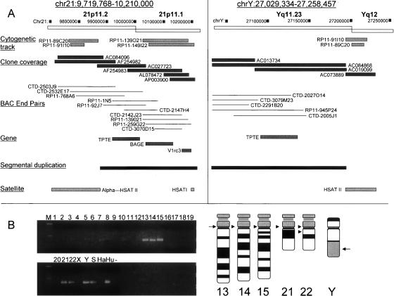
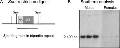
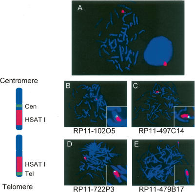
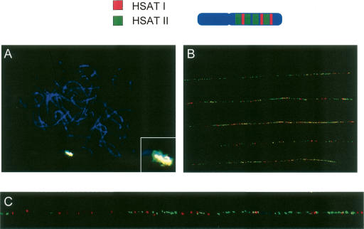
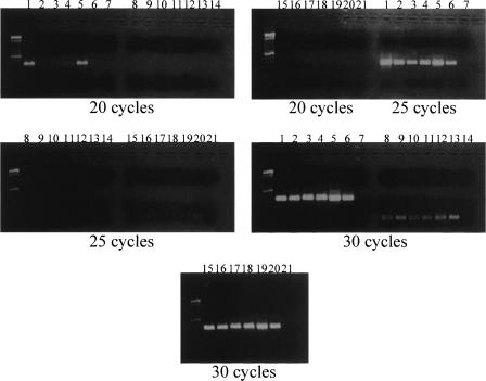
Low copy repeats (LCRs; segmental duplications) constitute approximately 5% of the sequenced human genome. Nonallelic homologous recombination events between LCRs during meiosis can lead to chromosomal rearrangements responsible for many genomic disorders. The 22q11.2 region is susceptible to recurrent and nonrecurrent deletions, duplications as well as translocations that are mediated by LCRs termed LCR22s. One particular DNA structural element, a palindromic AT-rich repeat (PATRR) present within LCR22-3a, is responsible for translocations. Similar AT-rich repeats are present within the two largest LCR22s, LCR22-2 and LCR22-4. We provide direct sequence evidence that the AT-rich repeats have altered LCR22 organization during primate evolution. The AT-rich repeats are surrounded by a subtype of human satellite I (HSAT I), and an AluSc element, forming a 2.4-kb tripartite structure. Besides 22q11.2, FISH and PCR mapping localized the tripartite repeat within heterochromatic, unsequenced regions of the genome, including the pericentromeric regions of the acrocentric chromosomes and the heterochromatic portion of Yq12 in humans. The repeat is also present on autosomes but not on chromosome Y in other hominoid species, suggesting that it has duplicated on Yq12 after speciation of humans from its common ancestor. This demonstrates that AT-rich repeats have shaped or altered the structure of the genome during evolution.


Figures

Figure 1.
Figure 1.
LCR22s mediate deletions/duplications and translocations. The line spanning left to right, represents the q11 region of chromosome 22 (cen indicates centromere). The LCR22s mediate the 22q11.2 deletion syndrome (velo-cardio-facial/DiGeorge syndrome), the reciprocal duplication syndrome, as well as the known recurrent t(11;22) translocation and t(17;22) translocation. LCR22-2 and -4 are >99% identical and through homologous recombination mediate the deletion syndromes, as well as, the duplication disorders. LCR22-3a is involved in the deletion syndromes as well but is predominately involved in translocations.
Figure 2.
Figure 2.
Organization of HSAT I/Alu/AT-rich sequence in the LCR22s. The LCR22s are made of blocks or modules shown here in the block structures (LCR22-2 is 241,823 bp; LCR22-3 size is not determined; LCR22-4 is 245,258 bp; Babcock et al. 2003). The different colors represent the different gene portions the LCR contains and its organization: USP18, pink; BCR, blue; GGT2 (found in LCR22-2 and LCR22-4, while GGT1 is found in LCR22-8) and GGTLA1, yellow; IGSF3 (pseudogene; real gene found on chromosome 1p13.1), orange. The blocks found under the LCR blocks represent the known HSAT I elements. These HSAT I elements are found in both orientations and are associated with Alus and AT-rich sequence. The orientation is centromere to telomere of 22q11.2 from left to right. The numbers represent the percent identity between the HSAT I’s, using the most centromeric element as reference.
Figure 3.
Figure 3.
Breakpoint at end of LCR22-2 is in AT-rich sequence. The image above shows the distal end of LCR22–2 (boxed) on chromosome 22q11.2 present in the genomic clone, AC008103. The DGCR6 gene is adjacent to the LCR22. The repetitive element track, showing LINEs, SINEs, AT-rich repeats, and HSAT I elements, adapted from the UCSC Browser genome assembly (http://genome.ucsc.edu/) is shown. The precise end of LCR22-2 is located within an AT-rich repeat. The sequences at the breakpoint are shown, with the unique sequence to the right of the AT-rich repeat. The position of the breakpoint is boxed. The chromosomal region containing LCR22-4 is shown below the respective region of LCR22-2. The genomic clone, AP000552 spans the interval. Similarly, the repetitive sequences are shown below the clone, with the position of the breakpoint, as boxed. Data are consistent with a breakpoint in the AT-rich repeat, within LCR22-4, resulting in the formation of LCR22-2.
Figure 4.
Figure 4.
HSAT I/AT-rich repeat is found on the acrocentric chromosomes and Y. (A, left) A chromosome 21p11 pericentromeric island of overlapping clones contains the tripartite repeat element (listed as HSAT I in the satellite track). The genomic clones used for physical (Clone coverage track) and FISH mapping (Cytogenetic track) are indicated below the boxes representing the acrocentric pericentromeric interval on chromosome 21p11.2 and 21p11.1 as adapted from the UCSC Browser, March 2006 assembly. Three genes were identified as shown, with their exon-intron structure (Gene track). The segmental duplication tract was incorporated into the diagram, with satellite type shown. There are three classes of satellites in this region, α-satellites, present at the very 5′ end of the chromosome 21p11.1 region; HSAT II [(GAATG)n]; and HSAT I satellites. (Right) The chromosome Yq11.23–12 junction contains similar features as the 21p11 region. There is striking similarity between the clones mapping to 21p11 and the Yq11.23-q12 interval. The Yq11.23-q12 junction is shown depicting the genomic clones used for physical and FISH mapping as adapted from the UCSC Browser. The 3′ ends of BAC clones AC019099, AC084868, and AC073889 contain HSAT II simple satellite sequence. The contigs are connected to the Yq12 interval based upon FISH mapping with clones RP11-91I10 and RP11-89C20. Additional validation as to the presence of HSAT II is shown in Figure 7. The TPTE gene maps to the 21p11 and Yq11.23 regions as shown. The tripartite repeat is missing from the reference human sequence for this interval and it is now shown in A. (B) The HSAT I element maps to the acrocentric chromosomes. PCR was performed using DNA template from the hamster-human somatic cell hybrid panel (Coriell repositories) using HSAT I F/R primers. The first lane represents a 100-bp ladder, each number represents the individual human chromosome, including X and Y. Ha is hamster DNA, Hu is human DNA, S is GM06317 (chromosome Y hamster–human somatic cell hybrid), and − is the negative control. The PCR product from different chromosomes were 400 bp in size. The acrocentric chromosomes 13, 14, 15, 21, 22, and Y were amplified. An idiogram of the acrocentric chromosomes and chromosome Y, with arrows in the putative position where the HSAT I elements are present, is shown.
Figure 5.
Figure 5.
Genomic Southern blot hybridization shows a specific hybridization in males as compared to females. (A) SpeI restriction digestion of genomic DNA. SpeI cuts once per tripartite repeat (HSAT I, open box; AluSc, light gray box; AT-rich sequence, dark gray box). The line below the genomic interval depicts the 2.4 kb fragment. (B) Genomic DNA was isolated from human blood lymphocytes and digested with SpeI, separated on 0.8% agarose gel and hybridized with a α-32P-dCTP–labeled HSAT I probe. The HSAT I probe detects the 2.4 kb tripartite repeat, which is present in greater copy number in male DNA as compared to female DNA.
Figure 6.
Figure 6.
Painting of chromosome Yq12. (A) FISH mapping on DAPI-stained metaphase and interphase human male peripheral blood lymphocytes with the 400-bp cloned PCR product of HSAT I, shown here in red. A signal was detected on chromosome Y. In the interphase cell, the hybridization of the HSAT I probe occurs near the periphery of the nucleus. We had two-dimensional images. Thus, we could not be sure of the position in all the nuclei. We saw the peripheral location in >60%–70% of 100 nuclei examined in separate experiments. (B) Positioning of HSAT I on chromosome Y. FISH mapping on DAPI-stained metaphase human male peripheral lymphocytes showing the position of HSAT I on the Y chromosome. (B,C) The BACs RP11-102O5 and RP11-497C14 (green) map more centromeric on chromosome Y than HSAT I (red). (D,E) The BACs RP11-722P3 and RP11-479B17 (green) map more telomeric on chromosome Y than the HSAT I probe. On the left of the FISH mapping pictures are the ideograms orientating the probes on the chromosome. HSAT I is localized on chromosome Y in the q12 band.
Figure 7.
Figure 7.
HSAT I and HSAT II FISH mapping. (A) FISH mapping on metaphase human male peripheral lymphocytes stained with DAPI with HSAT II probe (green) and HSAT I (red) have an overlapping signal producing a yellow color. (B) Fiber-FISH on chromosome Y with HSAT I probe (red) and HSAT II (green), show more hybridization of HSAT II on the Yq12 region compared with HSAT I. There is no distinct pattern of hybridization of HSAT I and HSAT II; however, HSAT II hybridizes to a larger region of the Yq12 region. (C) An enlarged region of one of the DNA fiber strands from B.
Figure 8.
Figure 8.
HSAT I semi-quantitative PCR on hominoid DNA. HSAT I, beta-catenin (CTNNB1), and SMARCB1 (viral integrase 1; two control primer sets) were used to amplify human, chimpanzee, pygmy, bonobo, gorilla, and orangutan DNA and then were electrophoresed in a 1.5% agarose gel. The PCR was done with increasing cycles 20, 25, and 30. (Lanes 17) HSAT I PCR, (lanes 814) CTNNB1, (lanes 1521) SMARCB1. The lanes are human–chimpanzee–pygmy–bonobo–gorilla–orangutan–negative control for each PCR primer and each cycling condition. The control primers do not amplify at the low cycles very well. In the HSAT I PCR, human (1) and gorilla (5) have an increased amplification compared to the other species.

Similar articles

Cited by

References

    1. Babcock M., Pavlicek A., Spiteri E., Kashork C.D., Ioshikhes I., Shaffer L.G., Jurka J., Morrow B.E., Pavlicek A., Spiteri E., Kashork C.D., Ioshikhes I., Shaffer L.G., Jurka J., Morrow B.E., Spiteri E., Kashork C.D., Ioshikhes I., Shaffer L.G., Jurka J., Morrow B.E., Kashork C.D., Ioshikhes I., Shaffer L.G., Jurka J., Morrow B.E., Ioshikhes I., Shaffer L.G., Jurka J., Morrow B.E., Shaffer L.G., Jurka J., Morrow B.E., Jurka J., Morrow B.E., Morrow B.E. Shuffling of genes within low-copy repeats on 22q11 (LCR22) by Alu-mediated recombination events during evolution. Genome Res. 2003;13:2519–2532. - PMC - PubMed
    1. Bailey J.A., Gu Z., Clark R.A., Reinert K., Samonte R.V., Schwartz S., Adams M.D., Myers E.W., Li P.W., Eichler E.E., Gu Z., Clark R.A., Reinert K., Samonte R.V., Schwartz S., Adams M.D., Myers E.W., Li P.W., Eichler E.E., Clark R.A., Reinert K., Samonte R.V., Schwartz S., Adams M.D., Myers E.W., Li P.W., Eichler E.E., Reinert K., Samonte R.V., Schwartz S., Adams M.D., Myers E.W., Li P.W., Eichler E.E., Samonte R.V., Schwartz S., Adams M.D., Myers E.W., Li P.W., Eichler E.E., Schwartz S., Adams M.D., Myers E.W., Li P.W., Eichler E.E., Adams M.D., Myers E.W., Li P.W., Eichler E.E., Myers E.W., Li P.W., Eichler E.E., Li P.W., Eichler E.E., Eichler E.E. Recent segmental duplications in the human genome. Science. 2002;297:1003–1007. - PubMed
    1. Bailey J.A., Liu G., Eichler E.E., Liu G., Eichler E.E., Eichler E.E. An Alu transposition model for the origin and expansion of human segmental duplications. Am. J. Hum. Genet. 2003;73:823–834. - PMC - PubMed
    1. Bandyopadhyay R., McQuillan C., Page S.L., Choo K.H., Shaffer L.G., McQuillan C., Page S.L., Choo K.H., Shaffer L.G., Page S.L., Choo K.H., Shaffer L.G., Choo K.H., Shaffer L.G., Shaffer L.G. Identification and characterization of satellite III subfamilies to the acrocentric chromosomes. Chromosome Res. 2001;9:223–233. - PubMed
    1. Benson G. Tandem repeats finder: A program to analyze DNA sequences. Nucleic Acids Res. 1999;27:573–580. - PMC - PubMed

Publication types

Substances

LinkOut - more resources