Review
. 2007 Mar;12(1):83-97.
doi: 10.1007/s10911-007-9040-x.

Extracellular proteolysis in transgenic mouse models of breast cancer

Affiliations
Review

Extracellular proteolysis in transgenic mouse models of breast cancer

Kasper Almholt et al. J Mammary Gland Biol Neoplasia. 2007 Mar.

Abstract

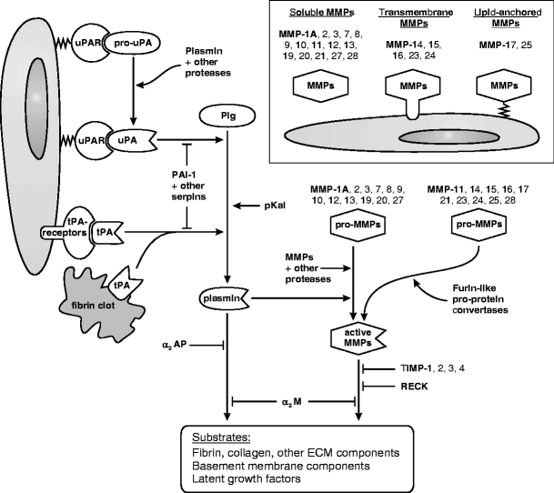
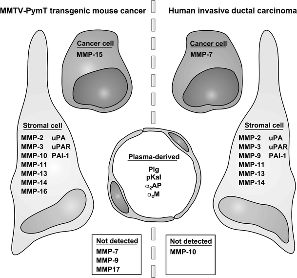
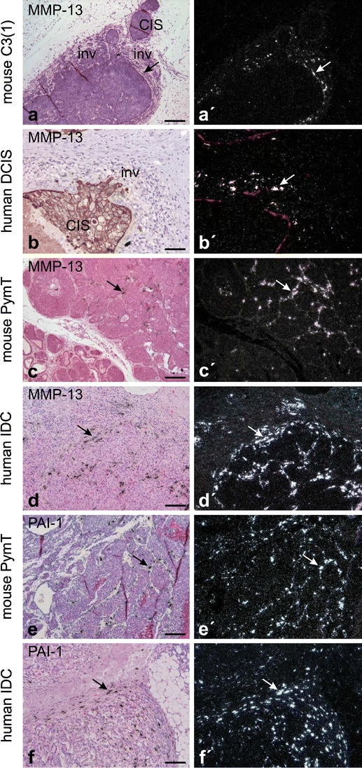
Growth and invasion of breast cancer require extracellular proteolysis in order to physically restructure the tissue microenvironment of the mammary gland. This pathological tissue remodeling process depends on a collaboration of epithelial and stromal cells. In fact, the majority of extracellular proteases are provided by stromal cells rather than cancer cells. This distinct expression pattern is seen in human breast cancers and also in transgenic mouse models of breast cancer. The similar expression patterns suggest that transgenic mouse models are ideally suited to study the role of extracellular proteases in cancer progression. Here we give a status report on protease intervention studies in transgenic models. These studies demonstrate that proteases are involved in all stages of breast cancer progression from carcinogenesis to metastasis. Transgenic models are now beginning to provide vital mechanistic insight that will allow us to combat breast cancer invasion and metastasis with new protease-targeted drugs.


Figures

Figure 1
Figure 1
The PA and MMP systems in the mouse. Plasmin and MMPs are two major sources of extracellular proteolysis. Plg is converted to the active protease plasmin by one of three Plg activators, uPA, tPA, and pKal. The generation of plasmin is enhanced by the binding of uPA to the cell surface receptor uPAR, and by the binding of tPA to coagulated fibrin or to cell surface proteins (annexin II [86] and CKAP4 [87]). The conversion of pro-uPA to active uPA may be initiated by several proteases, depending on the tissue in question. Additional pro-uPA is then activated by plasmin in a positive feed-back loop. The PA system is tightly controlled by several serine protease inhibitors (serpins); the primary and most specific inhibitors are the Plg activator inhibitor PAI-1 that targets uPA and tPA, and the plasmin inhibitor α2-antiplasmin (α2AP). Major substrates of plasmin include fibrin, fibronectin, laminin, and other ECM proteins, latent TGF-β and other growth factors, and pro-MMPs. The 22 different mouse MMPs are all extracellular, and are either soluble or membrane-anchored. Like the serine proteases of the PA system, the MMPs are also synthesized as inactive proforms that require proteolytic activation. Ten of the 22 pro-MMPs, including the membrane-anchored MMPs, have a furin cleavage site and may be activated by furin-like proprotein convertases before they are secreted. The remaining pro-MMPs are activated extracellularly, typically by plasmin or by other MMPs. The MMP system is counterbalanced by a group of four inhibitors, TIMPs, that have varying specificities for individual MMPs. In addition, the membrane-anchored MMP inhibitor RECK regulates a subgroup of MMPs including MMPs-2, -9, and -14 [88]. An excess of plasmin or MMP activity may ultimately be cleared by α2-macroglobulin (α2M) which is a non-specific protease scavenger. The MMPs are collectively able to degrade any component of the ECM. Important substrates for the MMPs as a group include the native fibrillar collagens and all four major components of the basement membrane: collagen type IV, laminin, nidogen/entactin, and heparan sulfate proteoglycans.
Figure 2
Figure 2
Expression of MMPs and PA components in breast tumors in humans and in MMTV-PymT transgenic mice. MMPs and PA components are expressed by cancer cells or stromal cells in breast tumors or are present as ubiquitous plasma-derived proteins. The predominant source of each mRNA/protein is illustrated for human ductal breast cancer and for the MMTV-PymT model in mice. Only those proteases and related components that have been analyzed in the MMTV-PymT model are included in the comparison. Only mRNA analyses are included except for uPAR, which was analyzed by immunohistochemistry in both species. The “Not detected” category includes proteases that were sporadically expressed in the tumors. The data for the MMTV-PymT model: uPA [31]; PAI-1 [45]; uPAR, MMPs-7, -9, -10 (unpublished data); MMPs-2, -3, -11, -13, -14, uPA, PAI-1 [43]; MMPs-14, -15, -16, -17 [46]. The data for invasive ductal breast cancer: uPA [47]; PAI-1 [48]; uPAR [52]; MMP-2 [49]; MMP-3 [50], MMP-7 [89]; MMP-9 [72]; MMP-10 [89]; MMP-11 [49]; MMP-13 [35]; MMP-14 [51].
Figure 3
Figure 3
Examples of protease expression in breast cancer from transgenic mice and humans. Expression of MMP-13 during early invasion in mouse C3(1)-SV40-T breast carcinoma (a) and in human ductal carcinoma in situ (DCIS) (b). MMP-13 is focally expressed in periductal fibroblast-like cells (arrows in a, a’, and b’) in areas with early cancer cell invasion (inv) and is not expressed in adjacent non-invasive areas with in situ carcinoma (CIS). Expression of MMP-13 in invasive MMTV-PymT carcinoma (c) and in human invasive ductal carcinoma (IDC) of the breast (d). Focal expression of MMP-13 is seen in fibroblast-like cells located in the stroma surrounding invasive cancer cells in both mouse and human breast cancers (arrows in c and d). Expression of PAI-1 in invasive MMTV-PymT carcinoma (e) and in human IDC of the breast (f). The expression of PAI-1 is seen in fibroblast-like cells located in the stroma surrounding invasive cancer cells in both the mouse and human breast cancers (arrows in e and f). All panels are in situ hybridization using 35S-labeled probes. Panels a, cf are counterstained with haematoxylin and eosin, whereas b is combined with pan-cytokeratin immunohistochemistry and (b’) with CK14 immunohistochemistry for myoepithelial cells [35]. Bars: a,c, 200 μm; b, 100 μm; df, 150 μm.

Similar articles

Cited by

References

    1. {'text': '', 'ref_index': 1, 'ids': [{'type': 'DOI', 'value': '10.1002/bies.20281', 'is_inner': False, 'url': 'https://doi.org/10.1002/bies.20281'}, {'type': 'PubMed', 'value': '16108064', 'is_inner': True, 'url': 'https://pubmed.ncbi.nlm.nih.gov/16108064/'}]}
    2. Green KA, Lund LR. ECM degrading proteases and tissue remodelling in the mammary gland. BioEssays 2005;27:894–903. - PubMed
    1. {'text': '', 'ref_index': 1, 'ids': [{'type': 'DOI', 'value': '10.1016/S0955-0674(98)80044-6', 'is_inner': False, 'url': 'https://doi.org/10.1016/s0955-0674(98)80044-6'}, {'type': 'PubMed', 'value': '9818179', 'is_inner': True, 'url': 'https://pubmed.ncbi.nlm.nih.gov/9818179/'}]}
    2. Johnsen M, Lund LR, Rømer J, Almholt K, Danø K. Cancer invasion and tissue remodeling: common themes in proteolytic matrix degradation. Curr Opin Cell Biol 1998;10:667–71. - PubMed
    1. {'text': '', 'ref_index': 1, 'ids': [{'type': 'DOI', 'value': '10.1007/s000180050497', 'is_inner': False, 'url': 'https://doi.org/10.1007/s000180050497'}, {'type': 'PMC', 'value': 'PMC11146824', 'is_inner': False, 'url': 'https://pmc.ncbi.nlm.nih.gov/articles/PMC11146824/'}, {'type': 'PubMed', 'value': '10949579', 'is_inner': True, 'url': 'https://pubmed.ncbi.nlm.nih.gov/10949579/'}]}
    2. Andreasen PA, Egelund R, Petersen HH. The plasminogen activation system in tumor growth, invasion, and metastasis. Cell Mol Life Sci 2000;57:25–40. - PMC - PubMed
    1. {'text': '', 'ref_index': 1, 'ids': [{'type': 'DOI', 'value': '10.1042/BST0300189', 'is_inner': False, 'url': 'https://doi.org/10.1042/bst0300189'}, {'type': 'PubMed', 'value': '12023849', 'is_inner': True, 'url': 'https://pubmed.ncbi.nlm.nih.gov/12023849/'}]}
    2. Bass R, Ellis V. Cellular mechanisms regulating non-haemostatic plasmin generation. Biochem Soc Trans 2002;30:189–94. - PubMed
    1. {'text': '', 'ref_index': 1, 'ids': [{'type': 'PubMed', 'value': '11003846', 'is_inner': True, 'url': 'https://pubmed.ncbi.nlm.nih.gov/11003846/'}]}
    2. Lund LR, Bjørn SF, Sternlicht MD, Nielsen BS, Solberg H, Usher PA, et al. Lactational competence and involution of the mouse mammary gland require plasminogen. Development 2000;127:4481–92. - PubMed

Publication types

Substances

LinkOut - more resources