

Roles of the innate immune system in mammary gland remodeling during involution
- PMID: 17286210
- PMCID: PMC2574498
- DOI: 10.1007/s10911-007-9036-6
Roles of the innate immune system in mammary gland remodeling during involution
Abstract
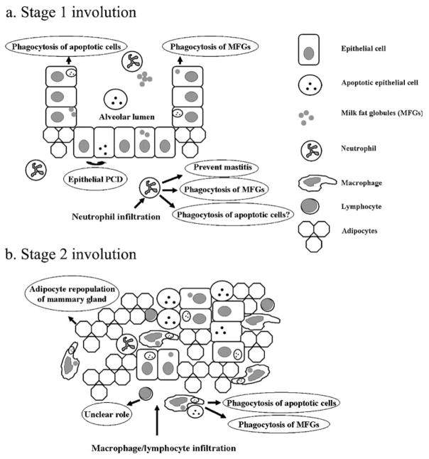
Mammary gland involution is a period of intensive tissue remodeling. Over the course of a relatively brief period, a large proportion of the mammary gland epithelium undergoes programmed cell death and is removed by phagocytes. In addition, the gland is cleared of residual milk fat globules as well as milk and adipocytes become the predominant cell type. The role of the immune system in this process has not been clearly defined. Professional phagocytes derived from the immune system can participate in the clearance of apoptotic and autophagic cells, the removal of residual milk components, and the prevention of mastitis during mammary gland involution. However, many of these functions can also be performed by non-professional phagocytes (e.g. mammary epithelial cells). This review will discuss the evidence that supports a role for innate immune cells in mammary gland remodeling during involution.
Figures
Similar articles
-
Impaired cell death and mammary gland involution in the absence of Dock1 and Rac1 signaling.Cell Death Dis. 2014 Aug 14;5(8):e1375. doi: 10.1038/cddis.2014.338. Cell Death Dis. 2014. PMID: 25118935 Free PMC article.
-
ATG proteins mediate efferocytosis and suppress inflammation in mammary involution.Autophagy. 2013 Apr;9(4):459-75. doi: 10.4161/auto.23164. Epub 2013 Feb 4. Autophagy. 2013. PMID: 23380905 Free PMC article.
-
Remodeling of Murine Mammary Adipose Tissue during Pregnancy, Lactation, and Involution.J Mammary Gland Biol Neoplasia. 2019 Sep;24(3):207-212. doi: 10.1007/s10911-019-09434-2. Epub 2019 Sep 12. J Mammary Gland Biol Neoplasia. 2019. PMID: 31512027 Free PMC article. Review.
-
Transcriptome profiling of the nonlactating mammary glands of dairy goats reveals the molecular genetic mechanism of mammary cell remodeling.J Dairy Sci. 2022 Jun;105(6):5238-5260. doi: 10.3168/jds.2021-21039. Epub 2022 Mar 26. J Dairy Sci. 2022. PMID: 35346464
-
Invited review: Accelerating mammary gland involution after drying-off in dairy cattle.J Dairy Sci. 2019 Aug;102(8):6701-6717. doi: 10.3168/jds.2019-16377. Epub 2019 Jun 13. J Dairy Sci. 2019. PMID: 31202662 Review.
Cited by
-
In-silico insights on the prognostic potential of immune cell infiltration patterns in the breast lobular epithelium.Sci Rep. 2016 Sep 23;6:33322. doi: 10.1038/srep33322. Sci Rep. 2016. PMID: 27659691 Free PMC article.
-
Leukocytes in mammary development and cancer.Cold Spring Harb Perspect Biol. 2011 Mar 1;3(3):a003285. doi: 10.1101/cshperspect.a003285. Cold Spring Harb Perspect Biol. 2011. PMID: 21123394 Free PMC article. Review.
-
Administration of Lactobacillus plantarum Lp62 to dam rats at the end of delivery and during lactation affects TGF-β1 level and nutritional milk composition, and body weight of pups.Eur J Nutr. 2019 Apr;58(3):1137-1146. doi: 10.1007/s00394-018-1628-y. Epub 2018 Feb 16. Eur J Nutr. 2019. PMID: 29453750
-
Efferocytosis creates a tumor microenvironment supportive of tumor survival and metastasis.Cancer Cell Microenviron. 2015;2(1):e666. doi: 10.14800/ccm.666. Cancer Cell Microenviron. 2015. PMID: 29264372 Free PMC article.
-
Semaphorin 7A Promotes Macrophage-Mediated Lymphatic Remodeling during Postpartum Mammary Gland Involution and in Breast Cancer.Cancer Res. 2018 Nov 15;78(22):6473-6485. doi: 10.1158/0008-5472.CAN-18-1642. Epub 2018 Sep 25. Cancer Res. 2018. PMID: 30254150 Free PMC article.
References
-
- Gouon-Evans V, Rothenberg ME, Pollard JW. Postnatal mammary gland development requires macrophages and eosinophils. Development. 2000;127(11):2269–82. - PubMed
-
- Marti A, Feng Z, Altermatt HJ, Jaggi R. Milk accumulation triggers apoptosis of mammary epithelial cells. Eur J Cell Biol. 1997;73(2):158–65. - PubMed
-
- Quarrie LH, Addey CV, Wilde CJ. Programmed cell death during mammary tissue involution induced by weaning, litter removal, and milk stasis. J Cell Physiol. 1996;168(3):559–69. - PubMed
Publication types
MeSH terms
Grants and funding
LinkOut - more resources
Full Text Sources
